

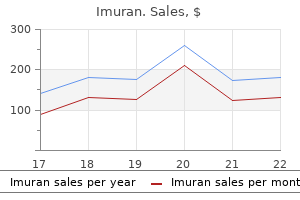
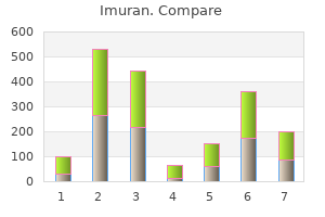
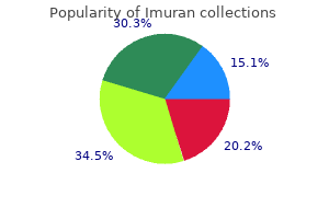
"Order imuran 50 mg, muscle relaxant with ibuprofen".
X. Kirk, MD
Clinical Director, University of Nevada, Reno School of Medicine
This is used either to maintain body potassium levels in patients receiving intravenous feeding, or to restore potassium levels in severely depleted patients. The main danger associated with intravenous potassium is hyperkalaemia, which can cause cardiac arrest. Potassium chloride has the dubious distinction of causing the highest frequency of fatal adverse reactions. Potassium chloride solution is infused at a maximum rate of 10 mmol/hour unless there is severe depletion, when 20 mmol/hour can be given with electrocardiographic monitoring. Potassium chloride for intravenous replacement should be dilute whenever possible. Potassium-retaining diuretics are better tolerated than oral potassium supplements. Replacement should be with appropriate volumes of crystalloid or blood in the case of haemorrhage. Hypokalaemia in untreated patients with hypertension is suggestive of mineralocorticoid excess. Severe hypokalaemia causes symptoms of fatigue and nocturia (because of loss of renal concentrating ability), and can cause dysrhythmias. Mild degrees of hypokalaemia (often associated with diuretic use) are generally well tolerated and of little clinical importance. Mild hypokalaemia is often unimportant, but severe hypokalaemia can cause dysrhythmias. However, if given by mouth it reacts with hydrochloric acid in the stomach to produce carbon dioxide, so it is poorly tolerated and not very effective. Instead, a citric acid/potassium citrate mixture can be used orally, as citrate is absorbed from the gut and metabolized via the tricarboxylic acid cycle with generation of bicarbonate. Potassium must be avoided in renal failure, as retention of potassium ions may cause hyperkalaemia. Calcium gluconate is a potentially life-saving emergency treatment in patients with dysrhythmias caused by hyperkalaemia (Chapter 32). Ion-exchange resin made of sodium or calcium polystyrene sulphonate removes potassium from the body in stool. The main adverse effect when resins are given chronically for patients with chronic renal failure is constipation, which can be avoided if the resins are suspended in a solution of sorbitol. Use Alkalinization of the urine is used to give symptomatic relief for the dysuria of cystitis and to prevent the formation of uric acid stones, especially in patients who are about to undergo cancer chemotherapy. The use of alkaline diuresis to increase urinary excretion of salicylate following overdose is discussed in Chapter 54. Mild hypokalaemia associated with thiazide or loop diuretics is common and seldom harmful per se. Where hypokalaemia is clinically important it can be corrected and/or prevented with K supplements or more conveniently with K -retaining diuretics. Emergency treatment of broad complex tachycardia caused by hyperkalaemia includes i. Haemodialysis or haemofiltration is frequently indicated in acute hyperkalaemic emergencies. When unstable detrusor contraction is responsible, drug treatment to reduce bladder activity is of limited use, combined with pelvic floor exercises and bladder training. Antimuscarinic drugs, such as oxybutynin, have a high incidence of antimuscarinic side effects. These may be minimized by starting with a low dose and by slow release formulation. Symptoms of benign prostatic hypertrophy may be improved by a 5reductase inhibitor.
The fluid passes from the lateral ventricles into the third ventricle through the interventricular foramina. The circulation is aided by the arterial pulsations of the choroid plexuses and by the cilia on the ependymal cells lining the ventricles. From the fourth ventricle, the fluid passes slowly through the median aperture and the lateral foramina of the lateral recesses of the fourth ventricle and enters the subarachnoid space. The fluid then moves through the cerebellomedullary cistern and pontine cisterns and flows superiorly through the tentorial notch of the tentorium cerebelli to reach the inferior surface of the cerebrum. Microvilli Ependymal epithelium of choroid plexus Cilia Tight junction Basement membrane Endothelium of blood capillary Blood Cavity of ventricle filled with cerebrospinal fluid Figure 16-16 Microscopic structure of the choroid plexus showing the path taken by fluids in the formation of cerebrospinal fluid. The dashed line indicates the course taken by fluid within the cavities of the central nervous system. It then moves superiorly over the lateral aspect of each cerebral hemisphere, assisted by the pulsations of the cerebral arteries. Some of the cerebrospinal fluid moves inferiorly in the subarachnoid space around the spinal cord and cauda equina. Here,the fluid is at a dead end,and its further circulation relies on the pulsations of the spinal arteries and the movements of the vertebral column, respiration, coughing, and the changing of the positions of the body. The cerebrospinal fluid not only bathes the ependymal and pial surfaces of the brain and spinal cord but also penetrates the nervous tissue along the blood vessels. Some of the cerebrospinal fluid probably is absorbed directly into the veins in the subarachnoid space, and some possibly escapes through the perineural lymph vessels of the cranial and spinal nerves. Because the production of cerebrospinal fluid from the choroid plexuses is constant, the rate of absorption of cerebrospinal fluid through the arachnoid villi controls the cerebrospinal fluid pressure. The arachnoid villi tend to be grouped together to form elevations known as arachnoid granulations. The arachnoid granulations increase in number and size with age and tend to become calcified with advanced age. The absorption of cerebrospinal fluid into the venous sinuses occurs when the cerebrospinal fluid pressure exceeds the venous pressure in the sinus. Electron-microscopic studies of the arachnoid villi indicate that fine tubules lined with endothelium permit a direct flow of fluid from the subarachnoid space into the lumen of the venous sinuses. Extensions of the Subarachnoid Space A sleeve of the subarachnoid space extends around the optic nerve to the back of the eyeball. The central artery and vein of the retina cross this extension of the subarachnoid space to enter the optic nerve, and they may be compressed in patients with raised cerebrospinal fluid pressure. The subarachnoid space also extends around the arteries and veins of the brain and spinal cord at points where they penetrate the nervous tissue. The pia mater, however, quickly fuses with the outer coat of the blood vessel below the surface of the brain and spinal cord,thus closing off the subarachnoid space. B: Magnified view of an arachnoid granulation showing the path taken by the cerebrospinal fluid into the venous system. Blood-Brain Barrier the experiments of Paul Ehrlich in 1882 showed that living animals injected intravascularly with vital dyes, such as trypan blue, demonstrated staining of all the tissues of the body except the brain and spinal cord. Later, it was demonstrated that although most of the brain is not stained after the intravenous injection of trypan blue, the following areas do in fact become stained: the pineal gland, the posterior lobe of the pituitary,the tuber cinereum,the wall of the optic recess, and the vascular area postrema1 at the lower end of the fourth ventricle. The permeability of the blood-brain barrier is inversely related to the size of the molecules and directly related to their lipid solubility. The barrier is almost impermeable to plasma proteins and other large organic molecules. Compounds with molecular weights of about 60,000 and higher remain Area of the medulla on the floor of the fourth ventricle just rostral to the opening into the central canal.
Whilst the advice and information in this book are believed to be true and accurate at the date of going to press, neither the authors nor the publisher can accept any legal responsibility or liability for any errors or omissions that may be made. In particular, (but without limiting the generality of the preceding disclaimer) every effort has been made to check drug dosages; however it is still possible that errors have been missed. Furthermore, dosage schedules are constantly being revised and new side-effects recognized. He was also aware of the need for a high quality textbook in clinical pharmacology that could also be used by nurses, pharmacists, pharmacology science students and doctors preparing for higher qualifications. It is interesting to follow in all the editions of the book, for example, how the treatment of tumours has progressed. John Trounce was pleased to see his textbook (and his subject) in the expert hands of Professor Ritter and his colleagues. Clinicians of all specialties prescribe drugs on a daily basis, and this is both one of the most useful but also one of the most dangerous activities of our professional lives. Understanding the principles of clinical pharmacology is the basis of safe and effective therapeutic practice, which is why this subject forms an increasingly important part of the medical curriculum. This textbook is addressed primarily to medical students and junior doctors of all specialties, but also to other professionals who increasingly prescribe medicines (including pharmacists, nurses and some other allied professionals). Clinical pharmacology is a fast moving subject and the present edition has been completely revised and updated. It differs from the fourth edition in that it concentrates exclusively on aspects that students should know and understand, rather than including a lot of reference material. Another feature has been to include many new illustrations to aid in grasping mechanisms and principles. The first section deals with general principles including pharmacodynamics, pharmacokinetics and the various factors that modify drug disposition and drug interaction. Drug metabolism is approached from a practical viewpoint, with discussion of the exciting new concept of personalized medicine. Adverse drug reactions and the use of drugs at the extremes of age and in pregnancy are covered, and the introduction of new drugs is discussed from the viewpoint of students who will see many new treatments introduced during their professional careers. Many patients use herbal or other alternative medicines and there is a new chapter on this important topic. There is a chapter on gene and cell-based therapies, which are just beginning to enter clinical practice. The remaining sections of the book deal comprehensively with major systems (nervous, musculoskeletal, cardiovascular, respiratory, alimentary, renal, endocrine, blood, skin and eye) and with multi-system issues including treatment of infections, malignancies, immune disease, addiction and poisoning. Their expertise in many specialist areas has enabled us to emphasize those factors most relevant. For their input into this edition and/or the previous edition we are, in particular, grateful to Professor Roy Spector, Professor Alan Richens, Dr Anne Dornhorst, Dr Michael Isaac, Dr Terry Gibson, Dr Paul Glue, Dr Mark Kinirons, Dr Jonathan Barker, Dr Patricia McElhatton, Dr Robin Stott, Mr David Calver, Dr Jas Gill, Dr Bev Holt, Dr Zahid Khan, Dr Beverley Hunt, Dr Piotr Bajorek, Miss Susanna GilmourWhite, Dr Mark Edwards, Dr Michael Marsh, Mrs Joanna Tempowski. We would also like to thank Dr Peter Lloyd and Dr John Beadle for their assistance with figures. If they are well, they may nevertheless want to know how future problems can be prevented. Depending on the diagnosis, treatment may consist of reassurance, surgery or other interventions. Drugs are very often either the primary therapy or an adjunct to another modality. Sometimes contact with the doctor is initiated because of a public health measure. Consequently, doctors of nearly all specialties use drugs extensively, and need to understand the scientific basis on which therapeutic use is founded. Thousands of potent drugs have since been introduced, and pharmaceutical chemists continue to discover new and better drugs. With advances in genetics, cellular and molecular science, it is likely that progress will accelerate and huge changes in therapeutics are inevitable. Medical students and doctors in training therefore need to learn something of the principles of therapeutics, in order to prepare themselves to adapt to such change.
Generally, when these infections occur in older children or adults, they are benign. However, if the host is immunocompromised or if the immune system is not yet developed, such as in the neonate, clinical symptoms may be quite severe or even fatal. Congenital infections can have manifestations that are clinically apparent antenatally by ultrasonography or when the infant is born, whereas perinatal infections may not become clinically obvious until after the first few days or weeks of life. When congenital or perinatal infections are suspected, the diagnosis of each of the possible infectious agents should be considered separately and the appropriate most rapid diagnostic test requested in order to implement therapy as quickly as possible. These immunoglobulin G (IgG) antibodies are acquired by passive transmission to the fetus and merely reflect the maternal serostatus. Pathogen-specific IgM antibodies do reflect fetal/infant infection status but with variable sensitivity and specificity. The following discussion is divided by pathogen as to the usual timing of acquisition of infection (congenital or perinatal) and in approximate order of prevalence. A summary of the diagnostic evaluation for separate viral infections is shown in Table 48. It is a member of the herpesvirus family, is found only in humans, and derives its name from the histopathologic appearance of infected cells, which have abundant cytoplasm and both intranuclear and cytoplasmic inclusions. Primary infection (acute infection) is usually asymptomatic in older infants, children, and adults, but may manifest with mononucleosis-like symptoms, including a prolonged fever and a mild hepatitis. Forty percent or more of pregnant women in the United States are 588 Infectious Diseases 589 Table 48. The risk of transmission to the fetus as a function of gestational age is uncertain, but infection during early gestation likely carries a higher risk of severe fetal disease. Vertical transmission can occur at any time in gestation or in the perinatal period, and infants are usually asymptomatic, especially if born to women seropositive before pregnancy. Additionally, 10% of the asymptomatic neonates will develop significant sequelae in the first year of life. Clinical disease in congenital infection may present at birth, while both congenital and perinatal infection can manifest with symptoms later in infancy. Congenital early symptomatic disease can present as an acute fulminant infection involving multiple organ systems with as high as 30% mortality. Laboratory abnormalities include elevated hepatic transaminases and bilirubin levels (as much as half conjugated), anemia, and thrombocytopenia. Hyperbilirubinemia may be present at birth or develop over time and usually persists beyond the period of physiologic jaundice. A second early presentation includes infants who are symptomatic but without life-threatening complications. These calcifications may occur anywhere in the brain, but are classically found in the periventricular area. Asymptomatic congenital infection at birth in 5% to 15% of neonates can manifest as later disease in infancy. Abnormalities include developmental abnormalities, hearing loss, mental retardation, motor spasticity, and acquired microcephaly. Other problems that can be detected later in life include inguinal hernia and dental defects due to abnormal enamel production. Almost all term infants who are infected perinatally remain asymptomatic, especially if the infection arose from a mother with reactivated viral excretion. Radiographically, there is hyperinflation, diffusely increased pulmonary markings, thickened bronchial walls, and focal atelectasis. A small number of infants may have symptoms that are severe enough to require mechanical ventilation, and historically, approximately 3% of infants die if untreated. Long-term sequela includes recurrent pulmonary problems, including wheezing and, in some cases, repeated hospitalizations for respiratory distress. Hematologic abnormalities were also seen, including hemolysis, thrombocytopenia, and atypical lymphocytosis. Depending upon local laboratory specifications, the specimen is collected with a Dacron swab, inoculated into viral transport medium, and then inoculated into viral tissue culture medium containing a coverslip on which tissue culture cells have been grown and incubated.