

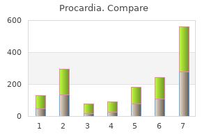
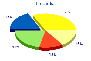
"Buy procardia on line, blood vessels map".
N. Bogir, M.A., M.D.
Clinical Director, Alpert Medical School at Brown University
Higher adherence to a detrimental pattern was associated with an increase in risk of 23 percent to 63 percent. These findings are also in agreement with the evidence linking dietary patterns and type 2 diabetes mellitus risk in populations of women who were not pregnant. Women of other races and ethnicities and those of lower socioeconomic status are underrepresented in this body of evidence. A major reason for grading this evidence as "limited" was the lack of adequately powered randomized controlled trials, few cohorts contributing to the observational studies, issues with risk of bias including self-reported For additional details on this body of evidence, visit: nesr. Chapter 2: Food, Beverage, and Nutrient Consumption During Pregnancy Not all components of the assessed dietary patterns were associated with all hypertensive disorders. Grade: Limited Evidence is insufficient to estimate the association between dietary patterns before and during pregnancy and risk of hypertensive disorders of pregnancy in minority women and those of lower socioeconomic status. An additional study showed an association between dietary patterns and blood pressure but not preeclampsia or gestational hypertension: o Dietary patterns characterized by higher intakes of vegetables, fruits, whole grains, nuts, legumes, fish, and vegetable oils were associated with a 30 percent to 42 percent decreased risk of hypertensive disorders and a 14 percent to 29 percent decreased risk of preeclampsia. One was associated with a 21 percent increased risk of preeclampsia and the other was associated with an increased risk of high blood pressure during pregnancy. Chapter 2: Food, Beverage, and Nutrient Consumption During Pregnancy o All but one of the studies were conducted outside the United States in samples that were predominantly White. The data were primarily observational, limiting the ability to draw any casual inferences. These patterns are higher in vegetables, fruits, nuts, legumes, fish and lower in added sugar, and red and processed meat. Eight of the 15 articles that assessed maternal dietary patterns using an index/score method 16 Scientific Report of the 2020 Dietary Guidelines Advisory Committee Part D. Studies had risk-of-bias issues, including exposure misclassification, self-reported outcomes, and selection bias. An eating occasion was defined as an ingestive event that is either energy yielding or non-energy yielding. This review identified 0 studies published between January 2000 and September 2019 that met the inclusion criteria for this systematic review. What is the relationship between dietary patterns consumed during pregnancy and gestational age at birth These protective dietary patterns are higher in vegetables, fruits, whole grains, nuts, legumes and seeds; and seafood (preterm birth only), and lower in red and processed meats and fried foods. Most of the research was conducted in healthy, Caucasian women with access to health care. Grade: Limited Evidence is insufficient to estimate the association between dietary patterns before pregnancy and gestational age at birth as well as the risk of preterm birth. Despite this variability, 5 of the 8 studies that assessed the relationship between dietary patterns during pregnancy and preterm birth found a statistically significant association. A sixth study found an association between dietary patterns during pregnancy and early preterm birth, but not preterm birth: o Highest adherence to a protective dietary pattern during pregnancy was associated with a preterm birth risk reduction of 9 percent to 90 percent. The fifth study showed an effect modification by parity: o Highest adherence to a protective dietary pattern during pregnancy was associated with a spontaneous preterm birth risk reduction of 15 percent to 45 percent. Generalizability of the included studies was limited to healthy White women who have access to health care. Women of other races and ethnicities and those with lower socioeconomic status are underrepresented in this body of evidence. Only 2 studies were conducted in the United States and 1 was primarily conducted in adolescent girls. Although research is available, the ability to draw a conclusion is restricted by inconsistency in study findings, inadequate adjustment of birth weight for gestational age and sex, and variation in study design, dietary assessment methodology, and adjustment of key confounding factors. Grade: Grade Not Assignable Insufficient evidence exists to estimate the association between dietary patterns before pregnancy and birth weight outcomes.
There will be a master computer in the meeting room and to ensure smooth transition between speakers and appropriate technical support, the Organisers request that speakers do not connect their own laptop. Every speaker has to go to the Preview room beforehand to provide his/her PowerPoint presentation. If you have video files attached to your power point presentation, they must be in the following format. The presentation for an early morning session should be handed over the evening before. You will also be able to review your presentation and to verify that it has been transferred correctly to the network. The Poster exhibition will be accessible from Friday to Monday in the exhibition area of the Maison de la Chimie, Paris. In the dedicated session slot, the presenting author must be available next to his/her dedicated board to present and discuss the poster. Text, tables and drawings for figures should be large enough to be seen from a 2 meters distance. Illustrations should be used to convey important points; diagrams, graphs, bar charts, scatter grams, pie charts and photographs will enhance your presentation. Prepare your material beforehand so that it will fit neatly into the space available and can be easily attached to the board. There will be a poster helpdesk close to the poster area, where staff will be happy to assist you. If you can have your poster produced by your institution, the final effect is more professional. A review of the benefits and limitations of both germline and somatic testing for each condition will be provided as well as specific case reports to highlight unique testing approaches that can be utilized in several clinical scenarios. The individual conditions were delineated as distinctive syndromes based on their characteristic findings and include von Recklinghausen disease (now known as neurofibromatosis type 1), Noonan syndrome, cardio-facio-cutaneous syndrome, Costello syndrome and others. These syndromes share central nervous system, cardiac and skin abnormalities, relative macrocephaly and short stature. It can be difficult to differentiate between these conditions, particularly in young patients, because the presentation of each syndrome is variable and may be age dependent. It should be outweighed by expected benefits for both the index patient and at-risk relatives. Appropriate counselling and testing strategies that serve to minimize the potential harm of testing should be available. The primary underlying genetic cause may remain elusive in familial schwannomatosis, and testing may rely on indirect linkage methods. Conclusions: Preimplantation and prenatal screening and testing options are changing rapidly. It is important for obstetric providers and medical geneticists to have a working knowledge of these options and their limitations, as well as societal guidelines regarding these test options. Melanocytes are cells which synthesize melanin pigment and thus contribute to the skin color. In addition to skinfolds, small pigment spots often cover large skin areas especially in trunk. Pale spots in skin may be part of nevus anemicus, which is a congenital vascular anomaly of the skin. Some anemic nevi are visible only after elicitation of vasodilatation in surrounding skin by gentle friction, while some are detectable without friction. Juvenile xanthogranulomas are predominant in infancy: 40% to 70% appear during the first year of life and they also disappear during childhood. Glomus tumours are benign but painful neoplasms of the glomus body of fingers and toes. Glomus bodies are thermoregulatory shunts which regulate capillary blood flow according to temperature.
The antigens responsible for the vast majority of the immune response are located on the endothelium. Whenever possible, corneal graft surgery is limited to anterior lamellar keratoplasty to minimize the immunogenicity of the graft tissue and the likelihood of rejection. Both humoral and cellular mechanisms have been implicated in corneal graft rejection. Lymphocytes mediating rejection generally move inward from the periphery of the cornea, forming a "rejection line" that may be seen on the endothelium or the epithelium as they move centrally. The donor cornea becomes edematous as the endothelium becomes increasingly compromised. Late rejection of a corneal graft may occur weeks to months after surgery and may be antibody-mediated, since cytotoxic antibodies have been isolated from the serum of patients with a history of multiple graft reactions in vascularized corneal beds. These antibody reactions are complement-dependent and attract polymorphonuclear leukocytes, which can form dense rings in the cornea at the sites of maximum deposition of immune complexes. Treatment the mainstay of the treatment of corneal graft reactions is intensive topical corticosteroid therapy. Due to its lack of regenerative capability, endothelial damage is likely to be irreversible, leading to graft failure such that endothelial rejection requires more aggressive and prolonged treatment. Systemic and topical cyclosporine or tacrolimus also are effective in preventing and treating graft rejection. Immunosuppressants these include antimetabolites (azathioprine, methotrexate, and mycophenolate mofetil), T-cell inhibitors (cyclosporine and tacrolimus), and alkylating agents (cyclophosphamide and chlorambucil). The treating physician must be familiar with the ocular and systemic side effects of these medications, which are discussed in Chapter 15. Examples of Biologic Response Modifiers (Biologics) for Ocular Disease Modes of Delivery In order to enhance anti-inflammatory effect on the eye and to minimize systemic side effects, alternative modes of delivery of these agents (apart from oral or intravenous) have also been studied. Intraocular methotrexate has shown vision improvement and 798 reduction of macular edema in uveitic patients. Sustained implants of immunosuppressants, as well as intraocular viral and nonviral gene therapies that deliver anticytokine agents, are being investigated in animal studies. Genetic predisposition to Stevens-Johnson syndrome with severe ocular surface complications. Symptoms are often nonspecific, and the usual examination techniques require modification. Development of the visual system is still occurring during the first decade of life, with the potential for amblyopia even in response to relatively mild ocular disease. Because the development of the eye often reflects organ and tissue development of the body as a whole, many congenital somatic defects are mirrored in the eye. Collaboration with pediatricians, neurologists, and other health workers is essential in managing these conditions. Similar collaboration is required in assessing the educational needs of any child with poor vision. Details of the embryology and the normal postnatal growth and development of the eye are discussed in Chapter 1. If any abnormality is identified, full ophthalmological assessment is required, for which the necessary instruments are hand light, loupe, direct and indirect ophthalmoscopes, and occasionally a portable slitlamp. Any congenital abnormality may be associated with nonocular abnormalities requiring further investigations. Pediatric Eye Examination Schedule 803 Vision Assessment of vision of the neonate is limited to observing the following response to a visual target, the most effective being a human face. Visual fixation and following movements can be demonstrated in most neonates; however, during the first 2 months of life, some do not demonstrate consistent fixation behavior and following (smooth pursuit) eye movements may be coarse and jerky. External Inspection the eyelids are inspected for growths, deformities, lid notches, and symmetric movement with opening and closing of the eyes.
Dark adaptation is often abnormal in retinal diseases characterized by rod photoreceptor dysfunction and impaired night vision. Fairbanks A et al: Ocular Ultrasound: A Quick Reference Guide for the OnCall Physician, 2016. Wang M: Corneal Topography in the Wavefront Era: A Guide for Clinical Application, 2nd ed. Using simple guidelines, patients requiring emergency or urgent ophthalmologic evaluation can be identified. Intensity and duration of pain, rapidity of onset and severity of visual loss (primarily assessed by visual acuity, which should be measured for each eye in all patients presenting with ophthalmic emergencies), gross appearance of the globe, and abnormalities on ophthalmoscopy are particularly important parameters. Excluding ocular and orbital trauma, which is covered in Chapter 19, this chapter reviews the common ophthalmic emergencies, for the most part grouped according to the predominant symptom. For each group, the section on triage highlights the features that are crucial during initial assessment such as on presentation to an emergency department. The section on clinical assessment emphasizes what is important during ophthalmologic evaluation. The management of the more common or important entities is then briefly discussed, principally to provide reference to discussion in other chapters. Conversely a few are at risk 136 of rapid progression within a few hours or days to severe visual impairment, even blindness, such as from acute angle-closure glaucoma, intraocular infection (endophthalmitis), bacterial, viral, amebic, or fungal corneal infection, acute uveitis, or scleritis. Triage (See Differential Diagnosis of Common Causes of the Inflamed Eye on Inside Front Cover) Emergency or urgent ophthalmologic evaluation should be arranged for any patient with acute red eye and a history within the past few weeks of intraocular surgery, which predisposes to endophthalmitis; contact lens wear, which predisposes to corneal infection; recent or distant history of corneal transplantation because of the possibility of graft rejection; previous episodes of acute uveitis or scleritis; or systemic diseases predisposing to uveitis or scleritis, such as ankylosing spondylitis and rheumatoid arthritis. In acutely ill patients, particularly those with sepsis or requiring prolonged intravenous cannulation such as in intensive therapy units or for parenteral nutrition, an acute red eye may be due to bacterial or fungal endophthalmitis. Ocular involvement in toxic epidermal necrolysis, Stevens-Johnson syndrome, or erythema multiforme requires urgent ophthalmologic assessment. Pain, rather than discomfort, should be regarded as inconsistent with conjunctivitis, episcleritis, or blepharitis. It is suggestive of keratitis, intraocular or scleral inflammation, or raised intraocular pressure, with the likelihood of a serious cause increasing with increasing severity. Associated nausea and vomiting are particularly suggestive of markedly raised intraocular pressure. Deep, boring pain, typically waking the patient at night, is characteristic of scleritis. Reduced vision, whether reported by the patient or identified by measurement of visual acuity, in the absence of a pre-existing explanation, should also be regarded as inconsistent with conjunctivitis, episcleritis, or blepharitis, and as with pain, the greater the severity, the greater is the likelihood of a serious cause. Severity of redness is not necessarily a guide to the seriousness of the underlying condition for example, despite its bright red appearance, subconjunctival hemorrhage is a benign entity. Distribution of redness can be helpful; predominance around the limbus (circumcorneal) is indicative of intraocular disease, diffuse redness involving the tarsal and bulbar conjunctiva is 137 indicative of conjunctivitis, focal or diffuse redness of the globe is consistent with episcleritis, and redness of the eyelid margins is indicative of blepharitis. Bluish redness (violaceous discoloration) of the globe, best identified in natural rather than artificial light, is characteristic of scleritis. Vesicles or ulcerations of the lids or periocular skin are typical of ophthalmic zoster (shingles) and less commonly varicella or primary herpes simplex virus infection. Conjunctivitis usually causes purulent, mucoid, or watery discharge, and allergic conjunctivitis typically causes itching. Profuse purulent discharge is characteristic of gonococcal conjunctivitis, which requires emergency treatment (see later in the chapter). A constricted pupil is suggestive of intraocular inflammation, typically due to anterior uveitis. Clinical Assessment Slitlamp examination facilitates assessment of distribution of redness; identification of conjunctival abnormalities, including examination of the superior tarsal conjunctiva following eversion of the upper eyelid; diagnosis of episcleritis and scleritis; characterization of corneal lesions; and detection of corneal keratic precipitates, anterior chamber flare and cells, and possibly hypopyon indicative of anterior chamber inflammation due to anterior uveitis, intraocular infection, or secondary to corneal inflammation, including infection. In cases of intraocular inflammation, dilated fundal examination is essential to determine whether there is involvement of the vitreous, retina, or choroid, which is important to diagnosis as well as assessment of severity (see Chapters 7 and 15). Management There are many causes of acute conjunctivitis, which in most cases is a benign, often self-limiting, condition (see Chapter 5). However, care needs to be exercised in neonates (ophthalmia neonatorum) (see Chapter 17) because of the possibility of infection with chlamydia or gonococcus, both of which are associated with nonocular disease that requires systemic therapy, or herpes simplex virus, which may be associated with encephalitis and requires hospitalization and parenteral antiviral therapy.