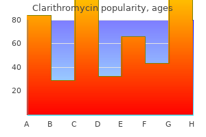
"Safe 500mg clarithromycin, gastritis symptoms lower back pain".
M. Mine-Boss, M.B. B.CH. B.A.O., M.B.B.Ch., Ph.D.
Associate Professor, Baylor College of Medicine
Additionally, myelography has the rare risk of causing pressure shifts leading to neurological decline if a complete subarachnoid spinal block is present. Radionucleotide Bone Scan Although radionucleotide bone scans cannot identify whether an epidural tumor is present, they are more sensitive than plain radiographs for detecting bone metastasis. Malignancies that do not trigger these physiological changes, such as multiple myeloma, are not detected. Additionally, numerous conditions in addition to cancer can demonstrate increased radionucleotide uptake, and the degree of thecal sac compression is unknown. Despite these shortcomings, one small retrospective study found that if both plain spinal radiographs and bone scans were negative in cancer patients with spinal symptoms, there was only a 2% risk of epidural spinal cord compression. First, a minimum of 50% of bone must be eroded before it is radiographically detectable. Second, plain films can only show bony changes and cannot address the issue of paraspinal tumor invading through the neural foramina or soft tissue impingement on the thecal sac or spinal cord. Third, metastatic involvement of multiple vertebra, as commonly occurs in breast and prostate cancer, may hide the clinically relevant lesion. The disk space, in contrast to infectious causes of vertebral collapse, usually remains intact. This is primarily because neither the epidural space nor the spinal cord can be clearly visualized. It is more sensitive, demonstrates the upper and lower limit of epidural metastases, shows the extraspinal extent of the tumor, and can demonstrate multiple epidural lesions. In one series, the probability of surviving 12 months was 73% for patients with restored or preserved ambulation following treatment but was only 9% for nonambulatory patients. As previously discussed, the most prevailing symptom of metastatic epidural spinal cord compression is pain, which can severely affect quality of life. Although some patients benefit from nonsteroidal anti-inflammatory medications, most require opiates for satisfactory pain relief. A good bowel regimen is required, as opiates, immobility, and autonomic dysfunction from a spinal lesion all contribute to constipation. Strict bed rest is not recommended for these patients, as they instinctively avoid activity that triggers discomfort. Those with small epidural deposits, no significant neurological dysfunction, and relative contraindications for glucocorticoids can safely begin radiation treatment without corticosteroid therapy. Corticosteroids work by decreasing spinal cord edema and may have an oncolytic effect on the tumor, especially lymphoma and breast cancer. Although the optimal dose fractionation scheme is not known, most radiation oncologists adhere to the standard schedule of 2500 to 3600 cGy in 10 to 15 fractions. This riskbenefit ratio depends on the time from first radiotherapy, initial dose, life expectancy, and probability of yielding worthwhile benefit. Metastatic epidural spinal cord compression from lesions originating from the breast or prostate may be treated with tamoxifen or androgen blockade, respectively. Cytotoxic agents may be efficacious in breast carcinoma or small cell lung cancer. Additionally, the rapidity with which the metastases respond to these agents depends on the predicated efficacy of the chemotherapy for each tumor type. By sectioning the denticulate ligaments, laminectomy decompresses the spinal cord, which allows it to move backward, avoiding anterior compression. However, when tumor causes vertebral body collapse, laminectomy may cause spinal instability and worsen neurological symptoms. This more aggressive technique, known as vertebral corpectomy, involves accessing the affected vertebral body and epidural space to completely debulk the tumor. This is followed by spinal column stabilization with either bone grafting or methylmethacrylate. After vertebral corpectomy and stabilization, 52% of patients had either complete or significantly improved neurological recovery, and 93% had good or excellent pain resolution. Metastatic spinal cord compression: occurrence, symptoms, clinical presentations and prognosis in 398 patients with spinal cord compression. Spinal epidural metastasis as the initial manifestation of malignancy: clinical features and diagnostic approach. The role of the vertebral venous system in metastases of cancer to the spinal column: experiments with tumor cell suspension in rats and rabbits.
The dendraxon of the pseudounipolar neuron may become convoluted to form a glomerulus. This nerve process then divides: One branch, a functional dendrite, passes to a receptor organ; the other, a functional axon, passes into the central nervous system. The perikarya of these pseudounipolar neurons do not receive synapses from other neurons. Autonomic ganglia consist of collections of perikarya of visceral efferent motor neurons and are located in swellings along the sympathetic chain or in the walls of organs supplied by the autonomic nervous system. Lipofuscin granules are more frequent in neurons of autonomic ganglia than in craniospinal ganglia. The ganglia of larger sympathetic chains are encapsulated by satellite cells, but a capsule may be absent around ganglia in the walls of the viscera. Unlike craniospinal ganglia, neurons of autonomic ganglia receive numerous synapses and are influenced by other neurons. Large nerve fibers are enclosed by a lipoprotein material called myelin; smaller nerve fibers may or may not be surrounded by myelin. Axons of peripheral nerves are enclosed by a sheath of flattened cells called Schwann cells. These cells are thin, attenuated cells with flattened, elongate nuclei located near the center of the cells. Their cytoplasm contains a small Golgi complex, a few scattered profiles of granular endoplasmic reticulum, scattered mitochondria, microtubules, microfilaments, and at times, numerous lysosomes. A continuous external (basal) lamina covers successive Schwann cells and the axonal surfaces of the nodes. The integrity of the external lamina is vital to regenerating axons and Schwann cells after injury. Schwann cells invest nerve fibers from near their beginnings almost to their terminations. The resulting neurilemma (sheath of Schwann) is continuous with the capsule of satellite cells that surrounds the perikarya of neurons in craniospinal ganglia. Schwann cells produce and maintain the myelin on nerve fibers of the peripheral nervous system. The neurilemmal and myelin sheaths are interrupted by small gaps at regular intervals along the extent of the nerve fiber. These interruptions represent regions of discontinuity between individual Schwann cells and are called nodes of Ranvier. The neurilemmal and myelin sheaths are made up of a series of small individual segments, each of which consists of the region between two consecutive nodes of Ranvier and makes up one internode or internodal segment. An internode represents the area occupied by a single Schwann cell and its myelin and is about 1 to 2 mm in length. Myelin is not a secretory product of Schwann cells but a mass of lipoprotein and glycolipid that includes galactocerebroside, which is abundant in myelin. Myelin results from a successive layering of the Schwann cell plasmalemma as it wraps around the axon. The wraps of cell membrane that form myelin are tightly bound together by specialized proteins. Myelin may appear homogeneous, but after some methods of preparation, it may be represented by a network of residual protein called neurokeratin. Ultrastructurally, myelin appears as a series of regular, repeating light and dark lines. The dark lines, called major dense lines, result from apposition of the inner surfaces of the Schwann cell plasmalemma. Po protein constitutes about 50% of the protein in peripheral nervous system myelin and aids in holding the myelin 108 together.
Hepatocytes are involved in the conversion of ammonium to urea, can perform gluconeogenesis (conversion of lipids and amino acids into glucose), synthesize cholesterol, as well as take up immunoglobulin A (IgA) and release it as a secretory from of IgA into the bile and ultimately into the lumen of the small intestine. Hepatocytes play a major role in the metabolism and storage of carbohydrates, fats, and proteins. A primary function of hepatocytes is to store carbohydrate in the form of glycogen. The surfaces of an individual hepatocyte either contact an adjacent liver cell or border on a perisinusoidal space. The plates of hepatocytes are 194 Hepatic glycogen functions to maintain normal blood glucose levels (70-100 mg/100 ml). During a high-carbohydrate meal as the blood glucose concentration begins to rise, a rapid pulse of insulin secretion occurs that results in an increase in the insulin glucagon ratio. The rise in insulin concentration suppresses hepatic glucose production and increases its uptake by insulindependent tissues. It is thought that a glucokinase, translocated from the nucleus to the cytosol during high glucose concentration catalyzes the phosphorylation of glucose and promotes glucose entry into hepatocytes. Insulin regulates glucokinase activity by both transcription and translocation in the hepatocytes. The storage form of glucose (glycogen) consists of glucose molecules linked by 1, 4 glycosidic bonds. Glycogen breakdown occurs during fasting and exercise that results in a decrease in the insulin glucagon ratio, and secretion of epinephrin. Liver glycogen is degraded by a hepatic glycogen phosphorylase to glucose-6-phosphate which in turn is catalyzed to free glucose by the enzyme, glucose-6-phosphatase, localized within the cisternae of the endoplasmic reticulum. Glucose-6phosphate is transported into the lumen of endoplasmic reticulum by a specific glucose-6phosphate transporter protein where it is hydrolyzed to glucose and phosphate by glucose-6phosphatase. Glucose is transported out of the endoplasmic reticulum and across the plasmalemma of the hepatocyte by glucose transport proteins. Hepatocytes also play a vital role in maintaining blood lipid levels; by the uptake of fatty acids and the esterification of fatty acids to triglycerides which occurs within the smooth endoplasmic reticulum. The triglycerides are then complexed to proteins within the Golgi complexes of hepatocytes to form a variety of lipoproteins. Amino acids are deaminated in the liver to produce urea which is excreted by the kidney. The liver also has functions in protein metabolism; synthesis of fibrinogen, prothrombin, and albumin; and storage of several vitamins (primarily A, D, B2, B3, B4 and B12). Enzymes associated with the smooth endoplasmic reticulum, on the other hand, are involved in the deactivation of some hormones, lipid soluble drugs, and toxins which is another important function of hepatocytes. Detoxified materials are excreted by hepatocytes into the bile and conducted by the biliary duct system to the intestinal tract for elimination. Bile is a complex exocrine alkaline secretion of the liver and contains ions, water, bicarbonate, bile acids (taurocholic and glycocholic), bile pigment (bilirubin glucuronide), phospholipids, cholesterol, lecithin, and neutral fats. Bile acids/salts act as emulsifying agents and are important in the breakdown of fat to micelles in the intestinal lumen during digestion. Most bile acids (about 80%) are reabsorbed in the ileum to be secreted once again into the bile. Bilirubin is produced in the spleen and liver as a result of the breakdown of the heme component of hemoglobin in damaged or old erythrocytes by macrophages at these locations. The bilirubin taken up by hepatocytes is then conjugated with glucuronic acid to form bilirubin glucuronide which is excreted into the bile. Bacterial action on this molecule after its entry into the intestinal tract converts it to urobilinogen. Of the urobilinogen formed, some is lost in the feces, some is absorbed and returned to the liver, and some is excreted by the kidneys. They are small and have thin walls, and their small lumina are surrounded by a low cuboidal epithelium that rests on a distinct basal lamina. The terminal ductules pass through the periportal limiting plate and empty into interlobular 195 bile ducts of the portal areas. The lumina of the bile ducts increase in diameter as they course toward the exterior, and the lining epithelium increases in height.
Autumn Blaze) and tissue cultured haplopappus (Haplopappus gracilis) were used in the experiment. Four different strains of haplopappus with different physiological and morphological characteristics were used: two aseptic clones, each generated from a single seedling; and two tissueculture-derived lines. The crew conducted a single inflight gas sampling procedure and a daily check of the experiment hardware to ensure proper function. Operations Preflight After the Shuttle landed, the specimens were recovered and photographed. The space-grown root tips were fixed and subsequently examined for rates of cell division and chromosomal aberrations. Selected individual specimens were successfully retrieved intact, grown through a full life cycle, and allowed to produce progeny for multigenerational postflight studies on the ground. Twenty-five shoots derived from aseptic suspension cultures of the monocot daylily and 75 plantlets of the dicot haplopappus had their roots severed prior to flight and were grown aseptically for four days in five Plant Growth Chambers on horticultural foam containing growth medium. The foam was washed to help prevent potentially detrimental components from leaching out of the foam and Results Root growth occurred randomly in all directions in space. In contrast, growth was uniformly positively gravitropic, so that roots grew in the direction of the gravity vector, in ground controls. Flight and groundcontrol plants produced equivalent amounts of tissue and maintained their characteristic root-production patterns. Capitulumderived plantlets are genetically disposed from their phenotype to have fewer roots because the energy is directed at producing the flower, whereas plantlets derived from seedlings would expect to have relatively normal root development because the seedling is directing development of the whole plant. The clonal root phenotype was not changed in space, at least for the short duration of the experiment. However, plantlets from both sources exhibited total root-production values that were 67 to 95 percent greater than those obtained in their ground-control counterparts. Microgravity may have brought about an altered moisture distribution pattern in the foam growth substrate, giving a more moist and thereby more favorable environment for root formation. Life Sciences Research Objectives Many space flight experiments have been conducted using plants. Most of these plants have shown poor vigor and have failed to reproduce successfully. In order to successfully provide bioregenerative life support (life support using plants for food, water, and waste removal), it is important to understand the reasons for these space flight effects. Programs, Missions, and Payloads 41 Life Sciences Payload Organisms Mouse-ear cress (Arabidopsis thaliana) plants were used in the experiment. This species is a flowering herb that is widely used for research in plant genetics because its genome size and short life cycle (45 days) make it an ideal candidate for gene mapping studies. Mouse-ear cress was also chosen because its small size allowed it to fit easily into the experiment hardware. Hardware the only experiment operation the crew performed was to conduct a daily check of the experiment hardware to ensure proper function. The plants reached the flowering stage on orbit and naturally occurring pollination of the flowers took place during the flight period. Operations Preflight After the Shuttle landed and the plants were recovered, reproductive leaf and root structures were preserved and subjected to morphological, histological, and biochemical analyses. Seeds were sown on agar 14 days prior to loading into the Plant Growth Chambers so that the plants would be developing flowers on orbit. Inflight Results There were striking differences between the flight-grown mouse-ear cress plants and the ground controls. Reproductive development was aborted at an early stage in the flight material in both male and female reproductive structures. Its objective was to study root growth and cell division in plants exposed to microgravity. The third experiment was designed to study the processes of cell wall formation and gene expression in microgravity. The seed production experiment used twelve 14-day-old mouse-ear cress plants (Arabidopsis thaliana).