

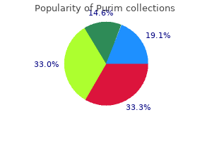
"Cheap purim, symptoms of anxiety".
B. Phil, M.B.A., M.B.B.S., M.H.S.
Clinical Director, Sanford School of Medicine of the University of South Dakota
The nonpolar alkyl chains are in the nonpolar phase; the polar carboxylate head groups are in the aqueous phase. Only in this way can amphiphilic detergents, through their hydrogen bonding with water and nonpolar interaction with a nonpolar (organic) phase or with air, maintain an orientation that ensures the lowest potential energy at an interface. A classic example of such behavior is given by soap, a mixture of alkali-metal salts of long-chain fatty acids. At a higher concentration, the molecules find it more energy efficient to "remove" their hydrophobic tails from the aqueous phase and let them interact with each other, thus forming a miniature "oil drop" or nonpolar phase, with the polar heads of the soap molecules in the bulk water. At a concentration that is characteristic for a given individual detergent, molecular aggregates, known as micelles, are formed. The concentration at which such micelles are formed is called the critical micellar concentration, and can be determined by measuring the light diffraction of the solution as a function of detergent concentration. When soap is dispersed in a nonpolar phase, inverted micelles are formed in which the nonpolar tails of the soap molecules interact with the bulk solvent while the hydrophilic heads interact with each other. This behavior of amphiphilic molecules explains how they can disperse nonpolar particles in water: the hydrocarbon tail of the amphiphile interacts with the particle, such as an oil droplet, dirt, or a lipoprotein membrane fragment, covers the particle, and then presents its hydrophilic head groups to the aqueous phase. When asked why he had stopped taking his phenytoin he stated that he had not, but had been taking the same dose for years. She stated that he took his daily dose of phenytoin every morning at breakfast and that she had witnessed his doing so, every day for the past six years. Upon questioning, it was discovered that this individual purchased his phenytoin in bottles of 1,000 capsules, in order to save money. He routinely stored this phenytoin in the basement of his home-a relatively damp and cool location-for months at a time; one week earlier he had started to use the capsules from one of these old stored bottles. When thirty "old" capsules from this recently opened bottle of 1000 capsules were weighed, all had masses in excess of 295 mg. When thirty capsules from a recently purchased supply of "new" phenytoin were weighed, all had masses less than 280 mg. An examination of an old capsule revealed that the contents were hardened and slightly discoloured. Subsequent analyses revealed that the phenytoin within the old capsules had become excessively hydrated from the ambient humidity of their storage conditions. The resulting hardened mass of drug material was less soluble within the gastrointestinal tract and was thus less bioavailable for absorption. Many factors can influence the bioavailability of a drug molecule following oral absorption. Damp storage conditions of the drug can cause increased molecular hydration with concomitant altered solubility. When a drug molecule is crystallized using different solvents or different conditions, the resulting change in crystal morphology can influence bioavailability and thus alter biological results. A rigorous control of molecular geometry and shape is crucial to the drug design process. Conformational isomerism is the process whereby a single molecule undergoes transitions from one shape to another; the physical properties of the molecule have not changed, merely the shape. Conformational isomerism is demonstrated by compounds in which the free rotation of atoms around chemical bonds is not significantly hindered. Rotation around torsional angles permits many different conformers (shapes) of a molecule. The concept and biophysical reality of "preferred" drug conformations and their potential role in receptor binding are currently important issues among drug designers. For aliphatic compounds, the well-known Newman projection is used to show the relative position of the substituents on two atoms connected to each other (as in ethane derivatives). When the trimethylammonium-ion and acetoxy functional groups are as far removed as possible, we speak of a fully staggered conformation (erroneously and confusingly also called a trans conformation).
The neuropsychology of facial expression: A review of the neurological and psychological mechanisms for producing facial expressions. Title: Clinical Neuroanatomy, 7th Edition Copyright ©2010 Lippincott Williams & Wilkins > Table of Contents > Chapter 12 - the Thalamus and Its Connections Chapter 12 the Thalamus and Its Connections A 61-year-old man with hypertension was seen in the emergency department, having apparently suffered a stroke. The patient was conscious and was unable to feel any sensation down the right side of his body. There was no evidence of paralysis on either side of the body, and the reflexes were normal. Three days later, the patient appeared to be improving, and there was evidence of return of sensation to the right side of his body. The patient, however, seemed to be excessively sensitive to testing for sensory loss. On light pinprick on the lateral side of the right leg, the patient suddenly shouted out because of excruciating burning pain, and he asked that the examination be stopped. Although the patient experienced very severe pain with the mildest stimulation, the threshold for pain sensitivity was raised, and the interval between applying the pinprick and the start of the pain was longer than normal; also, the pain persisted after the stimulus had been removed. Moreover, the patient volunteered the information that the pain appeared to be confined to the skin and did not involve deeper structures. Later, it was found that heat and cold stimulation excited the same degree of discomfort. The neurologist made the diagnosis of analgesia dolorosa or Roussy-Dejerine syndrome involving the left thalamus. This condition of thalamic overreaction is most commonly caused by infarction of the lateral nuclei of the thalamus due to hypertensive vascular disease or thrombosis. Understanding the functional role of the thalamus in the sensory system and knowing the central connections of the thalamus are necessary in making a diagnosis of thalamic disease. Chapter Objectives To review a very complex area of the nervous system and to emphasize that the thalamus lies at the center of many afferent and efferent neuronal loops to other parts of the nervous system the thalamus is situated at the rostral end of the brainstem and functions as an important relay and integrative station for information passing to all areas of the cerebral cortex, the basal ganglia, the hypothalamus, and the brainstem. There are two thalami, and one is situated on each side of the third ventricle. The posterior end is expanded to form the pulvinar, which overhangs the superior colliculus. The medial surface of the thalamus forms part of the lateral wall of the third ventricle and is usually connected to the opposite thalamus by a band of gray matter. Subdivisions of the Thalamus the thalamus is covered on its superior surface by a thin layer of white matter, called the stratum zonale. The gray matter of the thalamus is divided by a vertical sheet of white matter, the internal medullary lamina, into medial and lateral halves. The internal medullary lamina consists of nerve fibers that pass from one thalamic nucleus to another. The thalamus thus is subdivided into three main parts; the anterior part lies between the limbs of the Y, and the medial and lateral parts lie on the sides of the stem of the Y. Moreover, smaller nuclear groups are situated within the internal medullary lamina, and some are located on the medial and lateral surfaces of the thalamus. Anterior Part the anterior part of the thalamus contains the anterior thalamic nuclei. These anterior thalamic nuclei also receive reciprocal connections with the cingulate gyrus and hypothalamus. The function of the anterior thalamic nuclei is closely associated with that of the limbic system and is concerned with emotional tone and the mechanisms of recent memory. Medial Part the medial part of the thalamus contains the large dorsomedial nucleus and several smaller nuclei. The dorsomedial nucleus has two-way connections with the whole prefrontal cortex of the frontal lobe of the cerebral hemisphere. Lateral Part the nuclei are subdivided into a dorsal tier and a ventral tier.
Drug design for malaria is a research area with the capacity to blossom in coming years. Due to environmental changes such as global warming, the distribution of this disease may change, leading to malaria occurrences in the southern portions of North America and Europe. The search for antihelminthics has been somewhat less systematic than for other microbial pathogens. Most of these agents were discovered by traditional screening programs; their mechanisms of action at a molecular level are frequently unknown. It is thought to work by blocking microtubule synthesis in the nematode, subsequently impairing glucose uptake. Although its mechanism of action is basically unknown, it appears to somehow alter the surface structure of the helminth, rendering it more susceptible to destruction by the host defenses. Ivermectin appears to paralyze the nematode, which may lead to its death but which certainly facilitates its expulsion from the body. The drug appears to alter cholinesterase activity in the nematode, temporarily paralysing the adult worm and causing the worm to die. Niclosamide inhibits oxidative phosphorylation within the parasite, causing the cestode to be rapidly killed. This agent increases membrane permeability to Ca2+ in the nematode, causing paralysis, dislodgement, and death of the worm. Within the parasite, thiabendazole inhibits an enzyme called fumarate reductase, resulting in death of the worm. The infection may be diffuse (encephalitis of the brain) or localized (brain abscess). It may be caused by any of the infectious agents: meningitis may be caused by viral, bacterial, or fungal pathogens. The pathological consequences of infection may be immediate (meningococcal meningitis, causing rapid death) or delayed (syphilis, causing brain damage twenty years after the first infection). Similarly, the pathological ramifications of infections may be direct or indirect; for example, some researchers have speculated that a chlamydia infection of arteries may damage the arterial wall, causing atherosclerosis (leading to strokes and heart attacks) in later years. Clinically, pneumonia is characterized by fever, chills, shortness of breath, cough, and sputum production. The amount, viscosity, and color of the sputum are directly related to the type of organism causing the pneumonia. The infectious agents that cause pneumonia are many and varied, including many types of viruses, bacteria, and fungi. There is also a distinction between community-acquired and hospital-acquired (nosocomial) pneumonias; they may be caused by different microbes. The treatment of infectious diseases such as pneumonia is dictated more by the nature of the microbe than by the location of the infection. For example, a staphylococcal pneumonia is treated with the same antibacterial drugs as a staphylococcal infection elsewhere in the body. For example, an adenocarcinoma tumor of the lung is treated differently from adenocarcinomas elsewhere in the body. When given for a localized infection, antibiotics are not "smart bombs"; they are not tissue-specific and are widely distributed throughout the entirety of the body. Encephalitis is an inflammation, usually secondary to infection, of the substance of the brain. Whereas numerous viral, bacterial, fungal or parasitic agents are capable of producing the encephalitis syndrome, viral causes are the most frequent. The mortality and long-term morbidity rate of encephalitis is dependent upon the causative agent. The following two patients recently presented to an emergency room within minutes of each other. This 24-year-old male university student was brought to the emergency department at 1600 h by his roommate. Although he had been well the previous day, that morning he had complained of a fever, severe headache, severe neck and back stiffness, nausea, and vomiting. He was delirious and had neck rigidity with severe resistance to any attempt to passively flex his neck. Therapy was started immediately because of the life-threatening nature of the illness.
Syndromes
- Head MRI or CT scan of the brain
- Bowed legs and arms
- Intestinal symptoms include abdominal pain and diarrhea (which may be bloody).
- Galactosemia
- Protect your ears when you are exposed to loud noises. Wear protective ear plugs or earmuffs to protect against damage from loud equipment.
- Damage to nearby organs in the body
- Give your child any drugs your doctor told you to give your child with a small sip of water.
- Undescended testicle (cryptorchidism), or other testicular problems
The interval between the arachnoid and pia mater, the subarachnoid space, is filled with cerebrospinal fluid. The cerebrospinal fluid gives buoyancy to the brain and protects the nervous tissue from mechanical forces applied to the skull. The pia mater is a vascular membrane that closely invests and supports the brain and spinal cord. In lateral movements, the lateral surface of one hemisphere hits the side of the skull, and the medial surface of the opposite hemisphere hits the side of the falx cerebri. In superior movements, the superior surfaces of the cerebral hemispheres hit the vault of the skull, and the superior surface of the corpus callosum hits the sharp free edge of the falx cerebri; the superior surface of the cerebellum presses against the inferior surface of the tentorium cerebelli. Movements of the brain relative to the skull and dural septa may seriously injure the cranial nerves that are tethered as they pass through the various foramina. Furthermore, the fragile cortical veins that drain into the dural sinuses may be torn, resulting in severe subdural or subarachnoid hemorrhage. Intracranial Hemorrhage and the Meninges Epidural Hemorrhage Epidural hemorrhage results from injuries to the meningeal arteries or veins. The most common artery to be damaged is the anterior division of the middle meningeal artery. A comparatively minor blow to the side of the head, resulting in fracture of the skull in the region of the anterior-inferior portion of the parietal bone, may sever the artery. Arterial or venous injury is especially liable to occur if the vessels enter a bony canal in this region. Bleeding occurs and strips up the meningeal layer of dura from the internal surface of the skull. The intracranial pressure rises, and the enlarging blood clot exerts local pressure on the underlying motor area in the precentral gyrus. Blood also passes laterally through the fracture line to form a soft swelling under the temporalis muscle. The burr hole through the skull wall should be placed about 11/2 inches (4 cm) above the midpoint of the zygomatic arch. Subdural Hemorrhage Subdural hemorrhage results from tearing of the superior cerebral veins at their point of entrance into the superior sagittal sinus. The cause is usually a blow on the front or the back of the head, causing excessive anteroposterior displacement of the brain within the skull. In an epidural hemorrhage, the blood strips up the meningeal layer of the dura from the endosteal layer of dura (periosteum of the skull), producing a lens-shaped hyperdense collection of blood that compresses the brain and displaces the midline structures to the opposite side. The shape of the blood clot is determined by the adherence of the meningeal layer of dura to the periosteal layer of dura. In patients with subdural hematoma, the blood accumulates in the extensive potential space between the meningeal layer of dura and the arachnoid, producing a long crescent-shaped, hyperdense rim of blood that extends from anterior to posterior along the inner surface of the skull. With a large hematoma, the brain sulci are obliterated, and the midline structures are displaced to the opposite side. Subarachnoid and Cerebral Hemorrhages Subarachnoid and cerebral hemorrhages are described on page 484. Intracranial Hemorrhage in the Infant Intracranial hemorrhage may occur during birth and may result from excessive molding of the head. Excessive anteroposterior compression of the head often tears the anterior attachment of the falx cerebri from the tentorium cerebelli. Bleeding then takes place from the great cerebral veins, the straight sinus, or the inferior sagittal sinus. It follows that headaches are due to the stimulation of receptors outside the brain. Meningeal Headaches the dura mater receives its sensory nerve supply from the trigeminal and the first three cervical nerves. The dura above the tentorium is innervated by the trigeminal nerve, and the headache is referred to the forehead and face. The dura below the tentorium is innervated by the cervical nerves, and the headache is referred to the back of the head and neck. Meningitis, or inflammation of the meninges, causes severe headache over the entire head and back of the neck.