

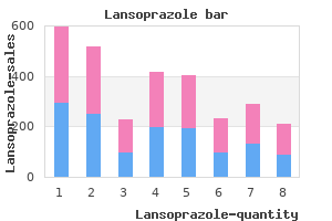
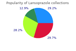
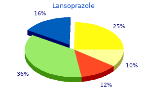
"Order lansoprazole pills in toronto, gastritis onions".
X. Hamid, M.S., Ph.D.
Co-Director, University of California, San Diego School of Medicine
In sympathetic ganglia, small amounts of acetylcholine stimulate postganglionic neurons and large amounts block transmission of impulses from preganglionic to postganglionic neurons. Consequently, these actions of acetylcholine are nicotinic actions and the receptors are nicotinic cholinergic receptors. Nicotinic receptors are subdivided into those found in muscle at neuromuscular junctions and those found in autonomic ganglia and the central nervous system. Both muscarinic and nicotinic acetylcholine receptors are found in large numbers in the brain. Each nicotinic cholinergic receptor is made up of five subunits that form a central channel which, when the receptor is activated, permits the passage of Na+ and other cations. Some of the receptors are homomeric-for example, those that contain five 7 subunits-but most are heteromeric. This opens the pore in the portion of the channel emnbedded in the lipid bilayer, and both K+ and Na+ flow through the open channel down their electrochemical gradient. The nicotinic cholinergic receptors in autonomic ganglia are heteromers that usually contain 3 subunits in combination with others, and the nicotinic receptors in the brain are made up of many other subunits. Many of the nicotinic cholinergic receptors in the brain are located presynaptically on glutamate-secreting axon terminals, and they facilitate the release of this transmitter. Some are located on structures other than neurons, and some seem to be free in the interstitial fluid, that is, they are perisynaptic in location. Each subunit has a binding site for acetylcholine, and when an acetylcholine molecule binds to each of them, they induce a confirmational change in the protein so that the channel opens. This increases the conductance of Na+ and other cations, and the resulting influx of Na+ produces a depolarizing potential. A prominent feature of neuronal nicotinic cholinergic receptors is their high permeability to Ca2+. Muscarinic cholinergic receptors are very different from nicotinic cholinergic receptors. The M4 receptor is found in pancreatic acinar and islet tissue, where it mediates increased secretion of pancreatic enzymes and insulin. Serotonergic Receptors the number of cloned and characterized serotonin receptors has increased rapidly. The transient hallucinations and other mental aberrations produced by this drug were discovered when the chemist who synthesized it inhaled some by accident. Its discovery called attention to the correlation between behavior and variations in brain serotonin content. It produces euphoria, but this is followed by difficulty in concentrating, depression, and, in monkeys, insomnia.
Diseases
- Hypothalamic hamartoblastoma syndrome
- Mesenteric panniculitis
- Thanatophoric dysplasia cloverleaf skull
- Optic atrophy polyneuropathy deafness
- Cinchonism
- Methylmalonic acidemia with homocystinuria
- Mesomelic dwarfism Nievergelt type
- Verloes David syndrome
The ampulla opens through the duodenal papilla, and its orifice is encircled by the sphincter of Oddi. Some individuals have an accessory pancreatic duct (duct of Santorini) that enters the duodenum more proximally. However, despite their substantial volume and fine control, gastric secretions are dispensable for the full digestion and absorption of a meal, with the exception of cobalamin absorption. This illustrates an important facet of gastrointestinal physiology, that digestive and absorptive capacity are markedly in excess of normal requirements. On the other hand, if gastric secretion is chronically reduced, individuals may display increased susceptibility to infections acquired via the oral route. Bile and intestinal juices are also neutral or alkaline, and these three secretions neutralize the gastric acid, raising the pH of the duodenal contents to 6. By the time the chyme reaches the jejunum, its pH is nearly neutral, but the intestinal contents are rarely alkaline. Bicarbonate ions are exported from the basolateral pole of the cell either by vesicular fusion or via a chloride/bicarbonate exchanger. It is therefore not surprising that the pancreas normally contains a trypsin inhibitor. Small amounts of pancreatic digestive enzymes normally leak into the circulation, but in acute pancreatitis, the circulating levels of the digestive enzymes rise markedly. Measurement of the plasma amylase or lipase concentration is therefore of value in diagnosing the disease. There is evidence for vagally mediated conditioned reflex secretion of pancreatic juice in response to the sight or smell of food. The bile acids contained therein are important in the digestion and absorption of fats. In addition, bile serves as a critical excretory fluid by which the body disposes of lipid soluble end products of metabolism as well as lipid soluble xenobiotics. Bile is also the only route by which the body can dispose of cholesterol-either in its native form, or following conversion to bile acids. In this chapter and the next, we will be concerned with the role of bile as a digestive fluid. In Chapter 29, a more general consideration of the transport and metabolic functions of the liver will be presented. Proteins and polypeptides Proteins and polypeptides Elastin, some other proteins Proteins and polypeptides Proteins and polypeptides Fat droplets Triglycerides Cholesteryl esters. Some of the components of the bile are reabRight hepatic duct Left hepatic duct sorbed in the intestine and then excreted again by the liver (enterohepatic circulation). The glucuronides of the bile pigments, bilirubin and biliverdin, are responsible for the golden yellow color of bile. The formation of these breakdown products of hemoglobin is discussed in detail in Chapter 29, and their excretion is discussed below. The bile acids secreted into the bile are conjugated to glycine or taurine, a derivative of cysteine. In common with vitamin D, cholesterol, a variety of steroid hormones, and the digitalis glycosides, the bile acids contain the steroid nucleus (see Chapter 22). The two principal (primary) bile acids formed in the liver are cholic acid and chenodeoxycholic acid. In the colon, bacteria convert cholic acid to deoxycholic acid and chenodeoxycholic acid to lithocholic acid. In addition, small quantities of ursodeoxycholic acid are formed from chenodeoxycholic acid. Water Bile salts Bile pigments Cholesterol Inorganic salts Fatty acids Phosphatidylcholine Fat Alkaline phosphatase 97. The bile salts have a number of important actions: they reduce surface tension and, in conjunction with phospholipids and monoglycerides, are responsible for the emulsification of fat preparatory to its digestion and absorption in the small intestine (see Chapter 27). They are amphipathic, that is, they have both hydrophilic and hydrophobic domains; one surface of the molecule is hydrophilic because the polar peptide bond and the carboxyl and hydroxyl groups are on that surface, whereas the other surface is hydrophobic. The numbers in the formula for cholic acid refer to the positions in the steroid ring.
A common design for spectroscopic studies is to compare the ratio of two spectral peak amplitudes between groups. We show that the correct propagation of this uncertainty improves statistical power. The method is able to statistically discriminates between signal and noise, and returns quantitatively bounded maps of rate constants of interest, such as $$$k { ext ightarrow ext}$$$. Whereas there was a trend of sex-related differences for few metabolites, no statistically significant differences were observed above the attained metabolite sensitivity threshold of 0. Reichenbach1 1 Medical Physics Group, Institute of Diagnostic and Interventional Radiology, Jena University Hospital - Friedrich Schiller University Jena, Jena, Germany, 2Institute for Physiotherapy, Jena University Hospital - Friedrich Schiller University Jena, Jena, Germany Due to specific training orientations athletes adapt with different metabolic responses to a given exercise. Different metabolic adaptations were shown within two muscles of different trained athletes. Data were analyzed utilizing linear discriminant in combination with principal component analysis. In addition, data from multiple time-points after injury demonstrate a return toward normal pattern and can be used to predict recovery and return-to-play times. Steatosis can be measured by magnetic resonance spectroscopy and imaging techniques non-invasively. At both points lipid and macromolecule magnetisation is excited from an approximately fully relaxed state and so this signal component is unchanged, thus the fitting model can be constrained. We show that this may be an advantageous strategy in comparison to other common techniques, reducing bias where fitting spectra with narrow linewidths. Relative inter- and intra-sequence variabilities with respect to B1 inhomogeneity and regional variation are reported. We optimize this sequence using simulations, and show its feasibility in phantoms and in vivo. We also explore the echo time modulation of the aspartate spin system using simulations and phantom experiments. Single exponential signal loss is assumed for metabolites, neglecting the possibility of non-linear decay at high b-values, as has been observed for water. Our goals are: (i) Characterize the metabolite signal decay versus b-value in human white matter to determine the non-linear region. This sequence has intrinsic water and lipid suppression properties due to the limited spectral bandwidth, so only prostate metabolites are measured. The scheme is based on transfer of voxels prescribed on an atlas to the subject images during the scanning session and allows fast and reliable placement of voxels for spectroscopy measurements. It is well-established that metabolite levels vary by brain region and voxel tissue composition. To address this problem, we developed and evaluated a novel and automated method of prescribing voxel placements at the time of scanning. Results demonstrated a significant improvement in prescribing accurate and reliable voxel placements between and within subjects compared to manual placement and published methods. We demonstrate an approach to measure transient field shifts arising from gradient-related eddy currents, and present a way to minimize these effects in order to restore correct editing. The relaxation-rate constant for the rapidly relaxing apparent water population is dominated by magnetization transfer and is insensitive pO2. Ram Manohar Lohia Institute of Medical Sciences, Lucknow, India, 2Centre of Biomedical Research, Lucknow, India, 3Department of Neurology, Dr. Ram Manohar Lohia Institute of Medical Sciences, Lucknow, India, 4Department of Microbiology, Dr. The study attempted to observe the metabolic variation in meningitis cases, negative controls and positive controls. The biomarkers identified were ketone bodies, amino acids, propylene glycol, citrate and creatine/creatinine. Chemometric analysis revealed a clear separation of samples from the different maturation days. Metabolite concentration evolutions could be followed and revealed strong and various metabolic alterations.
Define and give examples of direct inhibition, indirect inhibition, presynaptic inhibition, and postsynaptic inhibition. Describe the neuromuscular junction, and explain how action potentials in the motor neuron at the junction lead to contraction of the skeletal muscle. At chemical synapses, a synaptic cleft separates the terminal of the presynaptic cell from the postsynaptic cell. An impulse in the presynaptic axon causes secretion of a chemical that diffuses across the synaptic cleft and binds to receptors on the surface of the postsynaptic cell. This triggers events that open or close channels in the membrane of the postsynaptic cell. In electrical synapses, the membranes of the presynaptic and postsynaptic neurons come close together, and gap junctions form between the cells (see Chapter 2). Like the intercellular junctions in other tissues, these junctions form low-resistance bridges through which ions can pass with relative ease. There are also a few conjoint synapses in which transmission is both electrical and chemical. Regardless of the type of synapse, transmission is not a simple jumping of an action potential from the presynaptic to the postsynaptic cell. The effects of discharge at individual synaptic endings can be excitatory or inhibitory, and when the postsynaptic cell is a neuron, the summation of all the excitatory and inhibitory effects determines whether an action potential is generated. Thus, synaptic transmission is a complex process that permits the grading and adjustment of neural activity necessary for normal function. Because most synaptic transmission is chemical, consideration in this chapter is limited to chemical transmission unless otherwise specified. Transmission from nerve to muscle resembles chemical synaptic transmission from one neuron to another. The neuromuscular junction, the specialized area where a motor nerve terminates on a skeletal muscle fiber, is the site of a stereotyped transmission process. The contacts between autonomic neurons and smooth and cardiac muscle are less specialized, and transmission in these locations is a more diffuse process. Note that rough endoplasmic reticulum extends into the dendrites but not into the axon. Many different axons converge on the neuron, and their terminal boutons form axodendritic (4) and axosomatic (5) synapses. In some instances, the terminal branches of the axon of the presynaptic neuron form a basket or net around the soma of the postsynaptic cell (basket cells of the cerebellum and autonomic ganglia). In other locations, they intertwine with the dendrites of the postsynaptic cell (climbing fibers of the cerebellum) or end on the dendrites directly (apical dendrites of cortical pyramidal cells). It should be noted as well that synapses are dynamic structures, increasing and decreasing in complexity and number with use and experience. It has been calculated that in the cerebral cortex, 98% of the synapses are on dendrites and only 2% are on cell bodies. In the spinal cord, the proportion of endings on dendrites is less; there are about 8000 endings on the dendrites of a typical spinal neuron and about 2000 on the cell body, making the soma appear encrusted with endings. The postsynaptic density is an ordered complex of specific receptors, binding proteins, and enzymes induced by postsynaptic effects. Inside the presynaptic terminal are many mitochondria, as well as many membrane-enclosed vesicles, which contain neurotransmitters. Many presynaptic neurons terminate on dendritic spines, as shown at the top, but some also end directly on the shafts of dendrites.