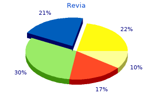
"Buy revia with a visa, medications pancreatitis".
T. Goose, M.A., M.D., M.P.H.
Deputy Director, Oklahoma State University Center for Health Sciences College of Osteopathic Medicine
The more thought put into this history taking, the more likely you are to not miss a significant injury. The time critical concept allows responders to not only identify life threatening injuries but to also establish the degree of urgency and, if necessary, indicate the speed of extrication or onward transport required. Once a time critical problem has been identified, solo responders should immediately stop their assessment to manage this problem. All time critical concerns must be constantly reassessed and reported clearly when care is handed over to the next team in the patients journey from roadside to critical care. If there are no signs of life under Respiratory or Circulation assessment, follow the dotted red arrows to initiate a Traumatic Cardiac Arrest Protocol Whilst some time critical of these may be treated on the spot, many will require transporting the patient and this should not be delayed. For example, getting a patient to the operating theatre for internal haemorrhage control must be considered a resuscitation measure, not necessarily definitive care. Attempting to "get them more stable" with intravenous fluids or blood transfusion is utterly misguided and will simply result in worsening blood loss, hypothermia and coagulopathy. Massive Haemorrhage the term massive external haemorrhage refers to a major bleed that is rapidly lifethreatening. Spurting arterial bleeds, blood soaked clothing, or pools of blood collecting on the floor should prompt a rapid assessment and immediate assessment and management. Massive haemorrhage must be aggressively addressed before any other casualty assessment takes place. The body has only a limited volume of circulating blood (5 litres), and once a large amount is lost (approximately 3 litres), as the Damage Control Resuscitation chapter will explain, it cannot be simply or effectively replaced with intravenous fluids or even blood transfusion. In a team, one member of the team can be assigned to massive haemorrhage management, whilst the rest of the team continue through the algorithm, working simultaneously. In a partially obstructed airway, the flow of air to the lungs is restricted, resulting in a harsh, high pitched noise known as stridor. The casualty may also cough and gag (which indicates that some air is passing around the obstruction) and speech will be severely impaired or even impossible. In a totally obstructed airway, no sounds of breathing effort can be heard, and no air is able to move in or out, despite good respiratory effort. If the airway is completely obstructed, the patient will lose consciousness rapidly. When an oxygen mask is used, the mask will fog every time the casualty breathes out through an open airway. This method of airway assessment allows the responder to count the respiratory rate and may be especially useful if access to the casualty is limited. Airway Airway compromise is the major cause of preventable deaths in prehospital trauma. Loss of the airway can deprive the brain and organs of vital oxygen and can lead to death in minutes. If the patient is totally unresponsive, or only responding to Pain, their airway is at risk and they are time critical. This assessment tool is particularly useful for a casualty trapped beyond your site or at a distance. Repiratory Rate Ask yourself, is this casualties respiratory rate normal for the environment you are in? An incredibly simple device, it should be the very first piece of monitoring placed on the patient. Whilst it may simply represent hypoperfusion from cold, it could indicate more serious respiratory or cardiovascular insult. Difficulty in Breathing Casualties with serious chest trauma often experience obvious distress or difficulty breathing. If the casualty is unable to complete full sentences, then this should raise concerns and justifies a more thorough assessment. Listen for any wheezing sounds or bubbling noises in the chest, which may indicate existing medical conditions such as asthma or heart failure. In trauma cases, these sounds may indicate serious chest injury and should be thoroughly assessed.
To identify this ischemic but not yet infarcted tissue virtually defines the goal of modern stroke treatment. Which of the sophisticated imaging techniques will contribute to improved clinical outcome is still to be determined, but certain ones, such as diffusionweighted imaging, have already proved invaluable in stroke work. First, all physicians have a role to play in the prevention of stroke by encouraging the reduction in risk factors such as hypertension and the identification of signs of potential stroke, such as transient ischemic attacks, atrial fibrillation, and carotid artery stenosis. Second, careful bedside clinical evaluation integrated with the newer testing methods mentioned above still provide the most promising approach to this category of disease. Finally, the last decade or two have witnessed a departure from the methodical clinicopathologic studies that have been the foundation of our understanding of cerebrovascular disease. Increasingly, randomized studies involving several hundred and even thousands of patients and conducted simultaneously in dozens of institutions have come to dominate investigative activity in this field. These multicenter trials have yielded highly valuable information about the natural history of a variety of cerebrovascular disorders, both symptomatic and asymptomatic. However, this approach suffers from a number of inherent weaknesses, the most important of which is that the homogenized data derived from an aggregate of patients may not be applicable to a specific case at hand. Moreover, many large studies show only marginal differences between treated and control groups. Each of these multicenter studies will therefore be critically appraised at appropriate points in the ensuing discussion. Since 1950, coincident with the introduction of effective treatment for hypertension, there has been a substantial reduction in the frequency of stroke. This was most apparent three decades ago, as treatment for high blood pressure became a public health focus. During this period, the incidence of coronary artery disease and malignant hypertension also fell significantly. In the last decade, according to the American Heart Association, the mortality rate from stroke has declined by 12 percent, but the total number of strokes may again be rising. Definition of Terms As discussed below, the term stroke is applied to a sudden focal neurologic syndrome, specifically the type due to cerebrovascular disease. The term cerebrovascular disease designates any abnormality of the brain resulting from a pathologic process of the blood vessels. Pathologic process is given an inclusive meaning- namely, occlusion of the lumen by embolus or thrombus, rupture of a vessel, an altered permeability of the vessel wall, or increased viscosity or other change in the quality of the blood flowing through the cerebral vessels. The vascular pathologic process may be considered not only in its grosser aspects- embolism, thrombosis, dissection, or rupture of a vessel- but also in terms of the more basic or primary disorder, i. Equal importance attaches to the secondary parenchymal changes in the brain resulting from the vascular lesion. These are of two main types- ischemia, with or without infarction, and hemorrhage- and unless one or the other occurs, the vascular lesion usually remains silent. The only exceptions to this statement are the local pressure effects of an aneurysm, vascular headache (migraine, hypertension, temporal arteritis), multiple small vessel disease with progressive encephalopathy (as in malignant hypertension or cerebral arteritis), and increased intracranial pressure (as occurs in hypertensive encephalopathy and venous sinus thrombosis). Also, persistent acute hypotension may cause ischemic necrosis in regions of brain between the vascular territories of cortical vessels, even without vascular occlusion. The many types of cerebrovascular diseases are listed in Table 34-1, and the predominant types during each period of life, in Table 34-2. Meningovascular syphilis, arteritis secondary to pyogenic and tuberculous meningitis, rare infective types (typhus, schistosomiasis, malaria, mucormycosis, etc. Connective tissue diseases (polyarteritis nodosa, lupus erythematosus), necrotizing arteritis. Wegener arteritis, temporal arteritis, Takayasu disease, granulomatous or giantcell arteritis of the aorta, and giant-cell granulomatous angiitis of cerebral arteries Cerebral thrombophlebitis: secondary to infection of ear, paranasal sinus, face, etc. Trauma and dissection of carotid and basilar arteries Amyloid angiopathy Dissecting aortic aneurysm Complications of arteriography Neurologic migraine with persistent deficit With tentorial, foramen magnum, and subfalcial herniations Miscellaneous types: fibromuscular dysplasia, with local dissection of carotid, middle cerebral, or vertebrobasilar artery, x-irradiation, unexplained middle cerebral infarction in closed head injury, pressure of unruptured saccular aneurysm, complication of oral contraceptives Undetermined cause in children and young adults: moyamoya disease and others Table 34-2 Cerebrovascular diseases characteristic of each age period 1.
If the sensory symptoms are localized to the head, the focus is in or adjacent to the lowest part of the convolution, near the sylvian fissure; if the symptoms are in the leg or foot, the upper part of the convolution, near the superior sagittal sinus or on the medial surface of the hemisphere, is involved. Lesions in or near the striate cortex of the occipital lobe usually produce elemental visual sensations of darkness or sparks and flashes of light, which may be stationary or moving and colorless or colored. According to Gowers, red is the most frequently reported color, followed by blue, green, and yellow. These images may be referred to the visual field on the side opposite of the lesion or may appear straight ahead. Curiously, a seizure arising in one occipital lobe may cause momentary blindness in both fields. More complex or formed visual hallucinations are usually due to a focus in the posterior part of the temporal lobe, near its junction with the occipital lobe, and may be associated with auditory hallucinations. The localizing value of visual auras has been confirmed recently by Bien and colleagues in a group of 20 surgically treated patients with intractable seizures. They found that elementary visual hallucinations and visual loss were typical of occipital lobe epilepsy but could also occur with seizure foci in the anteromedial temporal and occipitotemporal regions. Occasionally a patient with a focus in one superior temporal convolution will report a buzzing or roaring in the ears. A human voice, sometimes repeating unrecognizable words, or the sound of music, has been noted a few times with lesions in the more posterior part of one temporal lobe. Vertiginous sensations of a type suggesting a vestibular origin may on rare occasions be the first symptom of a seizure. The lesion is usually located in the superoposterior temporal region or the junction between parietal and temporal lobes. In one of the cases reported by Penfield and Jasper, a sensation of vertigo was evoked by stimulating the cortex at the junction of the parietal and occipital lobes. Occasionally with a temporal focus, the vertigo is followed by an auditory sensation. Giddiness, or light-headedness, is a frequent prelude to a seizure, but this symptom, as discussed in Chap. Gustatory hallucinations have also been recorded in proven cases of temporal lobe disease (see page 201) and with lesions of the insula and parietal operculum; salivation and a sensation of thirst may be associated. Electrical stimulation in the depths of the sylvian fissure, extending into the insular region, has produced peculiar sensations of taste. Vague and often indefinable visceral sensations arising in the thorax, epigastrium, and abdomen are among the most frequent of auras, as already indicated. Most often they have a temporal lobe origin, although in several such cases the seizure discharge has been localized to the upper bank of the sylvian fissure; in a few others, the focus was located in the upper or middle frontal gyrus or in the medial frontal area near the cingulate gyrus. Palpitation and acceleration of the pulse at the beginning of the attack have also been related to a temporal lobe focus. Complex Partial Seizures (Psychomotor Seizures, Temporal Lobe Seizures) these differ from the major generalized and absence seizures discussed above in that (1) the aura. Although it is difficult to enumerate all the psychic experiences that may occur during complex partial seizures, they may be categorized into a somewhat arbitrary hierarchy of illusions, hallucinations, dyscognitive states, and affective experiences. Objects or persons in the environment may shrink or recede into the distance, or they may enlarge (micropsia and macropsia), or perseverate as the head is moved (palinopsia). Hallucinations are most often visual or auditory, consisting of formed or unformed visual images, sounds, and voices; less frequently, they may be olfactory (usually unpleasant, unidentifiable sensations of smell), gustatory, or vertiginous. Emotional experiences, while less common, may be dramatic- sadness, loneliness, anger, happiness, and sexual excitement have all been recorded. Fear and anxiety are the most common affective experiences, while occasionally the patient describes a feeling of rage or intense anger as part of a complex partial seizure. Ictal fear may have no apparent connection to objective experience and is generally not related to the situation in which the patient finds himself during the seizure. Each of these subjective psychic states may constitute the entire seizure (simple partial seizure), or some combination may occur and proceed to a period of unresponsiveness. Patients call these "auras," but they represent electrical seizures and have the same significance as a motor convulsion.
As pointed out by Manford and colleagues, relatively few focal seizures can be localized precisely from clinical data alone. Somatosensory, Visual, and Other Types of Sensory Seizures Somatosensory seizures, either focal or "marching" to other parts of the body on one side, are nearly always indicative of a focus in or near the postrolandic convolution of the opposite cerebral hemisphere. Penfield and Kristiansen found the seizure focus in the postcentral or precentral convolution in 49 of 55 such cases. The sensory disorder is usually described as numbness, tingling, or a "pins-and-needles" feeling and occasionally as a sensation of crawling (formication), electricity, or movement of the part. In the majority of cases, the onset of the sensory seizure is in the lips, fingers, or toes, and the spread to adjacent parts of the body follows a pattern determined by sensory arrangements in the postcentral (postrolandic) convolution of the parietal lobe. The motor components of the seizure, if they occur, do so during the latter phase and take the form of automatisms such as lip-smacking, chewing or swallowing movements, salivation, fumbling of the hands, or shuffling of the feet. Patients may walk around in a daze or act inappropriately (undressing in public, speaking incoherently, etc. Certain complex acts that were initiated before the loss of consciousness- such as walking, chewing food, turning the pages of a book, or even driving- may continue. However, when asked a specific question or given a command, the patients are obviously out of contact with their surroundings. There may be no response at all, or the patient may look toward the examiner in a perplexed way or utter a few stereotyped phrases. In a very small number of patients with temporal lobe seizures (7 of 123 patients studied by Ebner et al), some degree of responsiveness (to simple questions and motor commands) is preserved in the presence of prominent automatisms such as lip-smacking and swallowing. Interestingly, in this small group of partially responsive patients, the seizures originate in the right temporal lobe. The patient, in a confused and irritable state, may resist or strike out at the examiner. Unprovoked assault or outbursts of intense rage or blind fury are very unusual; Currie and associates found such outbursts in only 16 of 666 patients (2. Penfield once commented that he had never observed a rage state as a result of temporal lobe stimulation. It is exceedingly unlikely that an organized violent act requiring several sequential steps in its performance, such as obtaining a gun and using it, could represent a temporal lobe seizure. Rarely, laughter may be the most striking feature of an automatism (gelastic epilepsy). A particular combination of gelastic seizures and precocious puberty has been traced to a hamartoma of the hypothalamus. Or the patient may walk repetitively in small circles (volvular epilepsy), run (epilepsia procursiva), or simply wander aimlessly, either as an ictal or postictal phenomenon (poriomania). These forms of seizure are actually more common with frontal lobe than with temporal lobe foci. Dystonic posturing of the arm and leg contralateral to the seizure focus is found to be a frequent accompaniment if sought- again, the origin is more often in the frontal than the temporal lobes, localizing particularly to the supplementary motor area. After the attack, the patient usually has no memory or only fragments of recall for what was said or done. Any type of complex partial seizures may proceed to other forms of secondary generalized seizures. The tendency to generalization holds true for all types of partial or focal epilepsy. The patient with temporal lobe seizures may exhibit only one of the foregoing manifestations of seizure activity or various combinations of them. In a series of 414 patients studied by Lennox, 43 percent displayed some of the motor changes; 32 percent, automatic behavior; and 25 percent, alterations in psychic function. Because of the frequent concurrence of these symptom complexes, he referred to them as the psychomotor triad. Probably the clinical pattern varies with the precise locality of the lesion and the direction and extent of spread of the electrical discharge. Because of their focal origin and complex symptomatology, all these types of seizures are best subsumed under the heading of complex partial seizures. This term is preferable to temporal lobe seizures, since typical complex partial seizures sometimes arise from a focus in the medial-orbital part of the frontal lobe.

The clinical effects of these several lesions are difficult to distinguish; there are some patients in whom it is impossible to state before operation whether the clot is epidural or subdural in location. A large acute clot causes a pineal shift as well as marked compression of one lateral ventricle; but if the clots are bilateral, there may be no shift and the ventricles appear symmetrical. Acute, rapidly evolving subdural hematomas are due to tearing of bridging veins, and symptoms are caused by compression of the brain by an expanding clot of fresh blood. Unlike epidural arterial hemorrhage, which is steadily progressive, venous bleeding is usually arrested by the rising intracranial pressure. Exceptionally, the subdural hematoma forms in the posterior fossa and gives rise to headache, vomiting, pupillary inequality, dysphagia, cranial nerve palsies, and, rarely, stiff neck and ataxia of the trunk and gait if the patient is well enough to be tested for these functions. A typical example is that of a child who has fallen from a bicycle or swing or has suffered some other hard blow to the head and was unconscious only momentarily. A few hours or a day later (exceptionally, with venous bleeding the interval may be several days or a week), headache of increasing severity develops, with vomiting, drowsiness, confusion, aphasia, seizures (which may be one-sided), hemiparesis with slightly increased tendon reflexes, and a Babinski sign. As coma develops, the hemiparesis may give way to bilateral spasticity of the limbs and Babinski signs. The pulse is often slow (below 60 beats per minute) and bounding, with a concomitant rise in systolic blood pressure (Cushing effect). Death, which is almost invariable if the clot is not removed surgically, comes at the end of a comatose period and is due to respiratory arrest. The visualization of a fracture line across the groove of the middle meningeal artery and knowledge of which side of the head was struck (the clot is usually on that side) are of aid in diagnosis and lateralization of the lesion. The surgical procedure consists of placement of burr holes in an emergency situation or, preferably, a craniotomy, drainage of the hematoma, and identification and ligation of the bleeding vessel. The operative results are excellent except in cases with extended fractures and laceration of the dural venous sinuses, in which the epidural hematoma may be bilateral rather than unilateral. If coma, bilateral Babinski signs, spasticity, or decerebrate rigidity supervene before operation, it usually means that displacement of central structures and crushing of the midbrain have already occurred; prognosis is then poor. There is always controversy about removing these in a patient who has no symp- Figure 35-9. Acute subdural hematoma over the right convexity, with substantial mass effect (displacement) of brain tissue but little edema. The head injury, especially in elderly persons and in those taking anticoagulant drugs, may have been trivial and may even have been forgotten. A period of weeks then follows when headaches (not invariable), light-headedness, slowness in thinking, apathy and drowsiness, unsteady gait, and occasionally a seizure or two are the main symptoms. The initial impression may be that the patient has a vascular lesion or brain tumor or is suffering from drug intoxication, a depressive illness, or Alzheimer disease. A gradual expansion of the hematoma is believed to cause the progression of symptoms. As with acute subdural hematoma, the disturbances of mentation and consciousness (drowsiness, inattentiveness, incoherence of thinking, and confusion) are more prominent than focal or lateralizing signs, and they may fluctuate. The focal signs usually consist of hemiparesis and rarely of an aphasic disturbance. Homonymous hemianopia is seldom observed, probably because the geniculocalcarine pathway is deep and not easily compressed; similarly, hemiplegia, i. An important feature of the hemiparesis from subdural hematoma, when it occurs, is that it may sometimes be ipsilateral to the clot, as a result of compression of the contralateral cerebral peduncle against the free edge of the tentorium by horizontal displacement of the midbrain (Kernohan-Woltman false localizing sign; see page 310). If the condition progresses, the patient becomes stuporous or comatose, but this course is often interrupted by striking fluctuations of awareness. With both large acute and chronic hematomas, dilatation of the ipsilateral pupil is a fairly reliable indicator of the side of the hematoma, though this sign may be misleading (occurring on the opposite side in 10 percent of cases, according to Pevehouse and coworkers). Convulsions are seen occasionally, most often in alcoholics or in patients with cerebral contusions, but they cannot be regarded as a cardinal sign of subdural hematoma. Rare cases of internuclear ophthalmoplegia and of chorea have been reported but have not occurred in our material. In infants and children, enlargement of the head, vomiting, and convulsions are prominent manifestations of subdural hematoma. It then becomes progressively hypodense (with respect to the cortex) over 2 to 6 weeks. The acute clot is hypointense on T2-weighted images, reflecting the presence of deoxyhemoglobin. Over the subsequent weeks, all image sequences show it as hyperintense as a result of methemoglobin formation.