

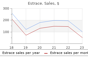
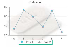
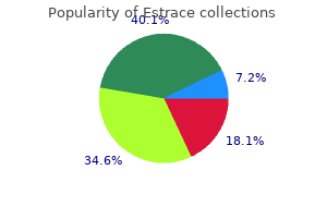
"Order estrace from india, breast cancer tattoo design".
A. Bram, MD
Vice Chair, Howard University College of Medicine
They constructed a Markov model from a societal perspective in order to perform a cost-effectiveness analysis. To date, the cost-effectiveness studies have focused mainly on other comparators than revascularization procedures or have made subsidiary comparisons. The medical history, the clinical state of the patient, and the outcome of cardiac angiography should determine which technique to use. Continued refinements in technology, increased knowledge regarding patient selection, and greater implanter experience are likely to further enhance safety and outcomes (3). Surgical Candidates and Preoperative Considerations for Neuromodulation Therapies Surgical candidacy reflects interplay between a surgically treatable condition, risk mitigation, and a benefit advantage. Agents that affect clotting pathways should be managed with the prescribing physician. Before surgery the patient should review with the anesthesiologist any and all perioperative concerns. The clinician should be alert for risk factors, such as poorly controlled diabetes, active infections, noncompliance, and psychological issues. Psychological Clearance for Neuromodulation Procedures Pain is, by definition, a sensory and emotional experience associated with or described in terms of actual or perceived tissue damage (1). The clinician may not recognize the impact of mood, depression, anxiety, or other negative affective states on the experience of pain. The challenge for the pain practitioner is to differentiate between the component that is biologically driven and the component that is emotionally driven or magnified. Anticoagulation Management No specific consensus documents have been published on neuromodulation and anticoagulation. Indeed, in a large retrospective survey comparing the outcomes of percutaneous leads vs. Training should include proper patient selection, contraindications, complication identification and management, and collaboration with colleagues in other pertinent disciplines. The implanter should be comfortable with troubleshooting during the implantation procedure and with methods and techniques to achieve proper stimulation while maintaining safety. The implanter should be able to recognize and treat hardware-related and biological complications and should be able to recognize the benefits and pitfalls of various commercial leads and lead types and their specific indications. Although implanters may choose to implant trial systems in the office setting, they should be able to obtain privileges to perform implantation in a Joint Commission-accredited hospital setting, properly certified surgical center, or similar facility. Additional information is available in a companion article that addresses the prevention and treatment of complications associated with neuromodulation (3). Accessing the epidural space either surgically or by needle placement is a common procedure with a very low incidence of bleeding, but any intervention demands vigilance for complications that might occur. A bridging method of anticoagulation from long-acting anticoagulants to short-acting anticoagulants may be implemented, as determined by the implanter and the prescribing physician. In existing case reports, detailed information is missing regarding the particular issues or difficulties encountered in the placement of the lead. Without these details, it is difficult to draw any firm conclusions regarding steps that need to be taken intraoperatively to prevent the occurrence of epidural hematoma. The evaluation should rule out intramedullary electrode placement, subdural hematoma, epidural hematoma, or spinal cord compression, along with any associated spinal canal stenosis. Treatment should be in accord with standard neurosurgical or surgical guidelines (3). Assessment of the trial outcome by the clinician includes evaluations of pain relief, improvement in patient function, associated treatment (in particular medication) utilization, and any complications of therapy. Preoperative Preparation Preoperative preparation for neuromodulation trials is patientspecific and disease-driven. This should, of course, occur with stable or, even better, reduced pain medications, in particular opioids, and with at least stable levels of daily activity. Ideally, objective data should be obtained by an independent observer like a physical therapist or rehabilitation specialist. The trial method to use may vary, depending on the indication, implanter, or patient. The rate of success of spinal cord stimulation is inversely related to time interval between onset of chronic pain syndrome and time of implantation. Neurosurgeons were the quickest to refer to an implant physician (average delay of 2. It should be remembered that these results of Kumar and Rizvi reflect the health-care system in Canada and might not apply to other national health-care systems.
Picture illustrating the joints that are most commonly affected by osteoarthritis. The muscles around a joint reduce the pressure on the joint and also stabilize it. It helps the bones in the joint to move smoothly over each other and it acts like a shock absorber that distributes the pressure from your body weight evenly throughout the joint. The joint is lined with a membrane called the synovium (represented by a black line). Bone Osteoarthritis is mostly found in people who are in their 50s and 60s or older. This does not mean that osteoarthritis is a disease due to old age, it is because the disease takes a long time to develop. There is often a genetic factor that contributes to osteoarthritis, so I recommend that you also read through the chapter on genetically inherited diseases on page 151. Secondary osteoarthritis is caused by trauma to a joint (such as rupture of a ligament) or a joint that is over used during competitive sports or a particular job. For example coal miners often develop osteoarthritis in the elbow joints, golfers in their big toe and foot ball players in the knees. Osteoarthritis can also result from a previous hip dislocation, a congenital deformity in the joint, abnormal bone structure (acromegaly) or a previous fracture in that bone. In secondary osteoarthritis there is damage or loss of the cartilage in the joint as a result of the injury. To compensate for this insult, the bone remodels the joint resulting in an altered pain free functioning joint. Sometimes, because of either an overwhelming injury or persistent injury on a chronic basis, this compensatory repair process fails, resulting in progressive tissue damage and secondary osteoarthritis. In the case of secondary osteoarthritis there is not necessarily a spiritual root behind it so you can just pray in faith for healing. If there is permanent damage to the bones, cartilage, ligaments or muscles you will have to operate in the gift of miracles. However the majority of people have primary osteoarthritis which involves none of the above scenarios. This is particularly true of the vertebra in the spine which can produce severe pain and disability. The joint damage is the Whatever goes on in your thought life, your brain converts into a physical reaction. As bitterness against yourself eats at you spiritually result of self bitterness: you in your thoughts, your body will eventually respond to this by in a sense are holding yourself guilty "eating itself". Interleukin 1 is a chemical that is part of the immune sysfor some issue in the past tem. When the cells of the immune system see these harmful substances in your blood stream or body tissues, the immune system kills or eliminates them, thus preventing you from becoming sick. However when you are attacking yourself spiritually with thoughts of self bitterness and guilt, the physical reaction via your brain results in interleukin 1 changing the biogenetic character of the cartilage cells. The cartilage cells release enzymes (called aggrecanase, collagenase and stromelysin) that degrade the structural components (called aggrecan and collagen) that make up the cartilage in your joints. This results in an increase in water concentration and swelling pressure in the cartilage that further damages the remaining scaffolding in the cartilage, making it vulnerable to load bearing injury. The cartilage is lost mainly at the area of the joint that takes most of the pressure of your body weight. Eventually the full thickness of the cartilage may be lost, resulting in the two bones in the joint grinding against each other. The changes in the cartilage encourage crystals (calcium pyrophosphate and apatite) to be deposited in the joint which can cause further irritation, inflammation and swelling. All the different parts that make up a joint (cartilage, bone, synovium, capsule, ligaments and muscle) depend on each other for health and function. Because the cartilage has decreased in thickness or has been lost altogether, the bone immediately below it increases in thickness (this is called sclerosis). This is caused by areas of bone that die as a result of the increased pressure in the bone because the weakened cartilage has failed in its load transmitting function.
The exercise of identifying bones is made pointless only if some routine is mindlessly mem orised simply to pass a possible examination question. However, if the bone is used to prompt a revision of its functions, then the exercise can be done with insight and purpose, and consequently becomes rewarding and easier. Moreover, if superficially similar bones have different functions or biomechanica1 needs, then subUe differences in structure can be sought and discovered, whereby individual bones can be recognised. Accordingly, both the structure of individua1 vertebrae and the structure of the entire lumbar spine should be considered. From above downwards, they increase in length and then decrease such that the L3 transverse process is usually the longest, and the transverse processes of L1 and L4 are usually the shortest. The other serial change in the lumbar spine is the orientation of the zygapophysial joints. Sagittally orientated joints are a feature of upper lumbar levels, while joints orientated. Examining this feature selVes l3 as a reminder of the compound role of the zygapophysial joints in resisting forward displacement and rotation, and the need at lower lumbar levels for stabilisation against forward displacement. From above downwards, the vertebral bodies tend to be sUghtly larger, and their transverse dimension tends to be relatively longer in proportion to their L4 anteroposterior dimension. The L4 vertebra forms a square, and the L5 vertebra forms a paralJelogram with its longer sides aligned horizontally. L4 will tend to have inferior articular processes orientated towards 45, and will have short transverse processes and a relatively wider body. Its transverse processes will be long and its articular processes will fall inside a rectangle. The L1 and L2 vertebrae remain with more sagittally orientated articular facets and articular processes that faU within trapezia. The only feature that may distinguish L1 from L2 is a better development of the mamiLiary and accessory processes on L1 and its shorter transverse processes. Apart from this, however, the upper two lumbar vertebrae may be indistinguishable. Its characteristic feature is the thickness of its transverse processes and their attachment along the whole length of the pedides as far as the vertebral body. Examining this feature serves to remind the student of the attachment of the powerful iliolumbar ligaments to the L5 transverse processes and their role in restraining the L5 vertebra. In tum, this is a reminder of the problem that L5 faces in staying in place on top of the sloping sacrum. There are no absolute features that enable the other four lumbar vertebrae to be distinguished but there are relative differences that reflect trends evident along the lumbar spine. Posterior longitudinal ligament posterior view L1-L4 (pedicles have been cut to show posterior aspect of the lumbar vertebral bodies) Lumbar Spine Innervation 5 1. Quadratus lumborum Multifidi lumborum Erector spinae Serratus posterior inferior Latissimus dorsi Thoracolumbar fascia 7. Variable histopathology of discovertebral lesion (spondylodiscitis) of ankylosing spondylitis. Delayed presentation and diagnosis of cervical spine injuries in long-standing ankylosing spondylitis. Magnetic resonance imaging examinations of the spine in patients with ankylosing spondylitis, before and after successful therapy with infliximab: evaluation of a new scoring system. Major reduction in spinal inflammation in patients with ankylosing spondylitis after treatment with infliximab: results of a multicenter, randomized, double-blind, placebo-controlled magnetic resonance imaging study. Clinical efficacy of etanercept versus sulfasalazine in ankylosing spondylitis subjects with peripheral joint involvement. Mortality, course of disease and prognosis of patients with ankylosing spondylitis. The contribution of genes outside the major histocompatibility complex to susceptibility to ankylosing spondylitis. Reactive arthritis: defined etiologies, emerging pathophysiology, and unresolved treatment. Comparison of sulfasalazine and placebo for the treatment of axial and peripheral articular manifestations of the seronegative spondylarthropathies: a Department of Veterans Affairs cooperative study.
Therefore, muscular action is transmitted to the vertebral body through the pedicles, which act as levers and thereby are subjected to a certain amount of bending. When a pedicle is bent downwards its upper wall is tensed while its lower wall is compressed. Similarly, if it is bent medially its outer wall is tensed while its inner wall is compressed. Through such combinations of tension and compression aJong opposite walls, the pedicle can resist bending forces applied to it. Consequently, there is no need for bone in the centre of a pedicle, which explains why the pedicle is hollow but surrounded by thick walls of bone. From opposite sides of the vertebral body, horizontal trabeculae sweep into the laminae and transverse processes. Within each process the extrinsic trabeculae from the vertebral body intersect with intrinsic trabeculae from the opposite A B Internal structure the trabecular structure of the vertebral body. Bundles of trabeculae sweep out of the vertebral body, through the pedicles, and into the articular processes, laminae and transverse processes. They reinforce these processes like internal buttresses, and are orientated to resist the forces and deformations that the processes habitually sustajn. The trabeculae of the spinous process are difficult to discern in detail, but seem to be anchored in the lamina and along the borders of the process. The other two are formed by the art iculation of the superior articular process of one vertebra with the inferior artic ular processes of the vertebra above. For example, the left L3-4 zygapophysial joint refers to the joint on the left, formed between the third and fourth lumbar vertebrae. The derivation relates to how, when two articulated vertebrae are viewed from the side, the articuJar processes appear to arch towards one another to form a bridge between the two vertebrae. It is popularised in the American literature, probably because it is conveniently short but it carries no formal endorsement and is essentially ambiguous. For example, in the thoracic spine, there are facets not only for the zygapophysial joints but also for the costovertebral joints and the costotransverse joints. Because the zygapophysial joints are located posteriorly, they are also known as the posterior intervertebral jOints. In fact, there is no formal name for the joint between the vertebral bodies, and difficulties arise if one seeks to refer to this joint. The only formal technical term for the joints between the vertebral bodies is the classification to which the joints belong. Moreover, if this system of nomenclature were adopted, to maintain consistency the zygapophysial joints would have to be known as the intervertebral diarthroses (see Table 1. It has been argued that this fashion is not consistent with the derivation of the word. The structure of the pars interarticularis of the lower lumbar vertebrae and its relation to the et iology of spondylolysis. Such a joint could adequately bear weight and would allow gUding movements between the two bodies. However, because of the Hatness of the vertebral surfaces, the joint would not allow the rocking movements that are necess. The first could be to introduce a curvature to the surfaces of the vertebral bodies. For example, the lower surface of a vertebral body could be curved (like the condyles of a femur). The upper vertebral body in an interbody joint could then roll forwards on the flat upper surface of the body below. Detailed structure of the intervertebral Constituents Microstructure Metabolism Functions of the disc Weight-bearing Movements 23 2. However, this Summary 26 adaptation would compromise the weight-bearing capacity and stability of the interbody joint. The bony surface in contact with the lower vertebra would be reduced, and there would be a strong tendency for the upper vertebra to roll backwards or forwards whenever a weight was applied to it. This adaptation, therefore, would be inappropriate if the weight-bearing capacity and stability of the interbody joint are to be preserved.