

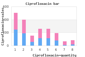
"Order ciprofloxacin without prescription, virus vaccines".
V. Arakos, M.A., Ph.D.
Co-Director, UTHealth John P. and Katherine G. McGovern Medical School
Electrocardiographicmarkersofabnormalleft ventricular wall motion in acute subarachnoid hemorrhage. Cardiac troponin elevation, cardiovascular morbidity, and outcome after subarachnoid hemorrhage. Plasma B-type natriuretic peptide levels are associated with early cardiac dysfunction after subarachnoid hemorrhage. Rebleeding of an aneurysm has devastating consequencesandisassociatedwithhighermortality. Moststudies show that the rerupture risk is highest in the first 24 hours and particularly within the first 6 hours after the ictus. Aneurysm size greater than 1 cm along with poor initial neurologicalpresentationandseizureatonsetwereriskfactorsfor rebleeding. Controversystillexistsregardinghypertensionasarisk factor versus a consequence of rebleeding. Reducing the risk of rebleeding before early aneurysm surgery: a possible role for antifibrinolytic therapy. Incidence and significance of early aneurysmal rebleeding before neurosurgical or neurological management. Interdisciplinary management results in 100 patients with ruptured and unruptured posterior circulation aneurysms. In a retrospective study, nicardipine was superior in controlling hypertension compared to labetolol. Esmolol and labetalol should be used if tachycardia needs to be treated along with hypertension. Pupillary involvement is almost pathognomonic for aneurysms, particularly if they are greater than or equal to 5 mm. In contrast, ischemic causes for third nerve palsies affect the central fibers, sparingthe pupil. Carotid artery aneurysms, particularly of the ophthalmic segment, but also anterior communicating and cavernous sinus aneurysms, can present with various visual field deficits such as bitemporal hemianopsia, homonymous hemianopsia, and quadrantanopsia. Ophthalmic aneurysmspresentinitiallywithaninferiornasalfieldcutbecause of the pressure on the overlying falciform ligament. Unruptured anterior communicating artery aneurysms can present with dementia, abulia, or pituitary dysfunctionfrommasseffect. Pupil-sparingthirdnervepalsywithpreoperative improvement from a posterior communicating artery aneurysm. Thestudyenrolledandfoundthat smaller aneurysms located in the anterior circulation had a lower ruptureriskcomparedtolargeraneurysmsandaneurysmslocatedin theposteriorcirculation. Neurogenicpulmonaryedemaisafairlycommon pulmonary complication and can be seen with any type of brain injury. The sympathetic surge causes capillary leakage regardless of the systemic blood pressure. Edema is caused by damage to capillary endothelium, which contains both alpha- and beta-adrenergic receptors. Accordingtotheblasttheory,thesystemicbloodpressure surgeshiftsbloodfromthesystemiccirculationtothelow-pressure pulmonary circulation, causing barotrauma to the capillary endothelium, damaging the alveolar membrane, and resulting in pulmonaryedema. Centrally distributed, bilateral infiltrates are seen on x-ray, associated with frothy blood-tinged sputum. The therapy is mainly supportive using mechanical ventilation as well as alphaantagonists like phentolamine, or beta-stimulating catecholamines to treat adrenergic-induced systemic vascular and pulmonary hypertension. Alternatively, some reports suggest dobutamine or dopamine to improve cardiac contractility. Several studies have shown that the incidence of cerebral vasospasm is increased with neurogenic pulmonaryedema,andsomestudieshaveshownahighermortality rate. These results are probably due to a higher initial Hunt and Hess grade and less aggressive hemodynamic resuscitation and hypertensive treatment of these patients due to aggravation of cardiac or respiratory failure with hypervolemic or euvolemic therapy. Itcanpresentasheadache,nausea,vomiting,anorexia,and lethargy, and, if untreated, these patients may rapidly develop cerebral edema and experience seizures and brain herniation. Bothcause hypotonichyponatremiawithelevatedurineosmolalitygreaterthan 200 mOsm/kg and urine Na greater than 25 mOsm/kg. This study, contrary to older literature, did not show an associationofhyponatremiawithvasospasmorpooroutcome.
Intracellular aggregates that accumulate elsewhere than in the lysosome are also a contributing factor in several types of neurodegeneration. Selective molecular binding activity to phosphorylated tau protein has been found in phosphorylated pSmad2 and pSmad3 proteins 1180 and curcumin, 1181 but numerous phosphorylation-dependent anti-tau antibodies. Phosphorylated Smad 2/3 colocalizes with phospho-tau inclusions in Pick disease, progressive supranuclear palsy, and corticobasal degeneration but not with alpha-synuclein inclusions in multiple system atrophy or dementia with Lewy bodies. An ultra-specific avian antibody to phosphorylated tau protein reveals a unique mechanism for phosphoepitope recognition. Passive immunization with Tau oligomer monoclonal antibody reverses tauopathy phenotypes without affecting hyperphosphorylated neurofibrillary tangles. Tau monoclonal antibody generation based on humanized yeast models: impact on Tau oligomerization and diagnostics. Harada R, Okamura N, Furumoto S, Furukawa K, Ishiki A, Tomita N, Tago T, Hiraoka K, Watanuki S, Shidahara M, Miyake M, Ishikawa Y, Matsuda R, Inami A, Yoshikawa T, Funaki Y, Iwata R, Tashiro M, Yanai K, Arai H, Kudo Y. Microbivores: Artificial mechanical phagocytes using digest and discharge protocol. Nanorobot activities near a particular tangle may need to be accompanied by the emission of small aliquots of an engineered enzyme designed to separate the neurofibrillary material from any cytoskeletal elements or other vital intracellular structures around which it might be wrapped or weakly bonded. Alternatively, the nanorobot can incorporate mechanisms and procedures designed to avoid damage to key intracellular structures while the tangles are being extracted and digested. Free-floating toxic tau oligomers are also extracted and safely digested by the nanorobots, possibly using binding sites derived from known molecular receptors that are specific to phosphorylated tau oligomers as described above, further reducing the rate of formation of new neurofibrillary tangles. Lipofuscin concentration in normal human brain early study 1211 obtained several as a function of age. How to create binding sites for such a heterogeneous material consisting of lipids, proteins, carbohydrates and a small amount of metals One approach is to find or create receptors for specific molecules found in lipofuscin membranes. For example, 186 different proteins 1213 were identified in the coatings of retinal epithelium lipofuscin granules, none of which were found in the purified lipofuscin when the coatings were chemically removed. A large number of stains are employed that preferentially bind to lipofuscin granules as an aid to visualization. Identification of proteins in lipofuscin using antibodies within the Human Protein Atlas;. Hydroxynonenal-generated crosslinking fluorophore accumulation in Alzheimer disease reveals a dichotomy of protein turnover. Specific lipofuscin staining as a novel biomarker to detect replicative and stress-induced senescence. Binding sites for sensing lipofuscin materials or lipofuscin granule membrane can be installed on the external recognition modules of tissue-mobile microbivore-class (Section 4. The effluent from these synthetic digestive processes would be mostly harmless free amino acids, fatty acids and carbohydrates, 1224 but any metal atoms present in the processed lipofuscin probably should be sequestered onboard the nanorobots. The microbivore-class devices would enter and exit the brain by any of several means described earlier in Section 4. It is not yet firmly established how quickly fresh lipofuscin would re-deposit in aging brain cells that have been completely cleared of the material. If the active deposition rate is similar to the ~1 mg/brain-yr rate for age-dependent accumulation, then decadal cleanouts may result in brain cells carrying only 10% of their natural lipofuscin load, reducing pathological effects to minimal levels. Medical bioremediation: prospects for the application of microbial catabolic diversity to aging and several major age-related diseases. In rat brain, -synuclein is highly expressed in the mitochondria in the olfactory bulb, hippocampus, striatum, and thalamus, where the cytosolic synuclein is also rich, whereas the cerebral cortex and cerebellum contains rich cytosolic synuclein but very low or even undetectable levels of mitochondrial -synuclein. The two morphological types are classical (brain stem) Lewy bodies and cortical Lewy bodies. Semi-quantitative analysis of alphasynuclein in subcellular pools of rat brain neurons: an immunogold electron microscopic study using a Cterminal specific monoclonal antibody. Cortical Lewy bodies are also composed of -synuclein fibrils, but are less defined and lack halos. In brain regions where Lewy bodies appear, such as the substantia nigra and locus coeruleus, up to 1-5 Lewy bodies per neuron have been observed.
Marcel Kinsbourne (1993) has postulated that this strong rightward orientation is less a function of right hemisphere dysfunction per se than a release of inhibition that lets the left hemisphere assert dominance in the presence of a now weakened right hemisphere. Perhaps some of the prime areas damaged, rendering the right hemisphere spatially ineffective, are locations within the right parietal lobe having to do with personal spatial frames of reference. Body position with respect to space is always egocentric, although people may have multiple frames with respect to bodies, heads, or position in relation to environment. Animal studies support the contention that there are distinct neuronal centers for these spatial frames within the right parietal cortex (for example, see Anderson, Snyder, Li, & Stricanne, 1993). Do these findings explain why the midline shift in neglect is nearly always to the right Bradshaw and Mattingly (1995) suggest that each hemisphere plays a specific role in spatial body position processing. According to this view, damage to the left parietal lobe produces no corresponding leftward shift because the spatial concerns of the left hemisphere are more feature oriented and language focused, resulting in a "no specialized spatial position" sense within the left parietal lobes. If right neglect does occur, they suggest, together with other investigators (such as Ogden, 1985), that the focus of the left hemisphere lesion would be anterior to the parietal lobes. Unfortunately, partly because of the rarity of occurrence, no research has explained the mechanisms of right-sided neglect. Understanding neglect is not only a problem of dominance and asymmetry, it is also an issue of conceptualizing the problem as a higher order network processing phenomenon. That unilateral neglect can occur with other nonparietal foci of damage is partial testament to this claim. We mentioned earlier that the region of the right inferior parietal lobe is the area most commonly damaged in cases of left unilateral neglect. As in other disorders discussed throughout this book, however, absence of function associated with a lesion does not necessarily imply that the lesioned area "contains" the function. Just as the hippocampus does not "contain" or store memory but is one of the most crucial links in memory processing and consolidation, the right inferior parietal lobe does not in itself contain "body mindfulness" but may be a crucial link. Neuroanatomically, left unilateral neglect also occurs with damage to a variety of subcortical structures, most notably the thalamus (see Bradshaw & Mattingly, 1995). It is reasonable to speculate that neglect results from a disconnection in higher order processing that involves the coordination of many second-order systems, such as visual processing, attention, memory, and possibly other systems. They are beyond the scope of this overview, and excellent reviews exist elsewhere (for example, see Bradshaw & Mattingly, 1995). Most models describe the process of body and hemispace cognition as including visual-perceptual processes, attention, and motor action. The parietal lobes control perceptual processing, the premotor and prefrontal cortices mediate exploratory-motor behavior, and the cingulate gyrus directs motivation. In turn, subcortical structures probably coordinate the orchestration of all three areas. The reticular formation directs arousal, and the thalamus (particularly the pulvinar of the thalamus) is postulated to focus and guide attention between spatial locations. The primary visual system, which serves as a "feature analyzer," builds to the higher order systems of object recognition and spatial localization. Disorders such as visual agnosia and neglect provide good examples of how each of these systems may malfunction. However, with vision, there is much reciprocal networking between parallel systems, so what appears hierarchical may not be entirely so. What is discussed in a bottom-up fashion may also be affected by top-down processing. Currently, brain science has uncovered much of the structure and many of the functions of the visual road map through the brain. However, much work needs to be done in understanding how the brain accommodates to varying visual experiences that represent the same precept.

Smith continued, "I do not have any concrete evidence to prove this, but it is my opinion. To establish eligibility for a consequential condition under Part E, you must prove by preponderance of the evidence that the diagnosed illness, in this case sleep apnea, occurred as a result of an accepted illness. This is established by a fullyrationalized medical report by a physician which shows the relationship between the claimed consequential condition and the accepted illness. The "at least as likely as not" standard cited in your request only pertains to causation determinations for primary illnesses, which is not at issue in your claim for a consequential illness. Finally, the request for reconsideration renewed your previous objections to the June 9, 2010 recommended decision of the Denver district office. The denial of your Part E claim for sleep apnea is final on the date of issuance of this denial of your request for reconsideration. Such a request must be in writing and must be made within 30 days of the date of issuance of this decision. In order to ensure that you receive an independent evaluation of the evidence, your request for reconsideration will be reviewed by a different Final Adjudication Branch hearing representative than that who issued the final decision. If your claim was denied because a cancer was not causally related to work-related exposure to radiation and you can identify either a change in the probability of causation guidelines, a change in the dose reconstruction methods or an addition of a class of employees to the Special Exposure Cohort, you may also request a reopening of your claim. Should you wish to receive this type of review; the district office will provide you with a determination. Please note, however, that such a determination does not change your eligibility for benefits or establish causation under the Act, and is not subject to further agency or judicial review. Except as provided above, all future correspondence, inquiries or telephone calls should be directed to the district office. The district office is directed to evaluate the new medical evidence in support of your claims, and issue a new Recommended Decision to address your eligibility under both Part B and Part E of the Act. The Director has delegated the authority to review and issue determinations for certain claims to the District Director having jurisdictional authority over the case. With regard to the claimed condition of colon cancer, you did not submit medical evidence to establish the diagnosis. As such, the district office issued four letters, dating from July of 2004 to December of 2004, requesting that you provide evidence to establish your diagnosis with colon cancer, and that the condition resulted from your occupational exposure to a toxic substance. The case is returned to the Cleveland District Office to proceed as outlined below. Subsequently, on May 1, 2006, the district office also issued a Recommended Decision to deny your claim for colon cancer under Part E; again citing insufficient evidence that you were diagnosed with the claimed illness. In a submission received by the district office on May 26, 2007, you provided a pathology report and additional medical records to confirm your diagnosis with colon cancer. Accordingly, the district office referred your case file to the Office of the Director for review and consideration of reopening claims under both Part B and Part E of the Act. This new evidence includes a pathology report dated January 16, 2002, confirming your diagnosis with colon cancer. Additionally, various medical reports and progress notes, ranging from 2002 to the present, establish your diagnosis and treatment for this illness. This new evidence invalidates the basis of the May 6, 2005 and June 10, 2006 Final Decisions denying your Part B and Part E claims. As such, it is necessary to vacate these prior Final Decisions so that the district office may proceed with a new examination of your eligibility under Part B and Part E for colon cancer. The May 6, 2005 and June 10, 2006 Final Decisions, respectively, denying your claim for colon cancer under Part B and Part E are vacated. The district office is directed to evaluate the new medical evidence in support of your claims and issue a new Recommended Decision to encompass your eligibility to benefits for the condition of colon cancer under both Part B and Part E of the Act. I have reviewed the objections and the evidence on file and I find that your case is not in posture for reopening at this time. The attached Denial of Reopening Request provides further explanation of why there is insufficient basis to warrant reopening.
Source of Infection and Mode of Transmission: Cyclosporiasis is acquired through ingestion of raw fruits and vegetables and contaminated water. A later study, in which 5,552 stool samples were collected from workers on raspberry farms in Guatemala, found infection rates of between 2. A study of the water in domestic containers in Egypt showed that 56% was contaminated with Giardia, 50% with Cryptosporidium, 12% with Blastocystis, 9% with Cyclospora, and 3% with microsporidia (Khalifa et al. Using microscopy and molecular biology techniques, Sturbaum (1998) identified Cyclospora oocysts in wastewater. They can be detected by staining, autofluorescence under ultraviolet light, phase contrast microscopy, or polymerase chain reaction (Ortega et al. The stains used most frequently (to make it easier to visualize the organisms and to differentiate them from yeasts) are trichrome stains, Ziehl-Neelsen, Giemsa, safranin with methylene blue, calcofluor white, and auramine phenol. Safranin has been found to be the most effective and appropriate stain for use in diagnostic laboratories (Negm, 1998). Control: Cyclosporiasis can be prevented by applying the classic measures for control of parasitoses transmitted via the fecal-oral route: washing foods that are eaten raw, boiling suspicious water, and washing hands before eating. Treatment of water contaminated with Giardia, Cryptosporidium, Blastocystis, Cyclospora, or microsporidia with chlorine at 4 or 8 parts per million (ppm) or with ozone at 1 ppm showed that ozone was more effective in destroying the parasites, but that it did not totally inactivate Cyclospora or Blastocystis (Khalifa et al. Cyclospora cayetanensis infections in Haiti: A common occurrence in the absence of watery diarrhea. Attempts to establish experimental Cyclospora cayetanensis infection in laboratory animals. Epidemiology and treatment of Cyclospora cayetanensis infection in Peruvian children. Isolation of Cryptosporidium parvum and Cyclospora cayetanensis from vegetables collected in markets of an endemic region in Peru. Etiology: the taxonomy of the species of the genus Giardia is still controversial. Although in the past many species were described and named according to the host in which they were found-for example, G. Although Lamblia was the original name given to the genus by Lambl when he first described it in 1859, Stiles changed it to Giardia in 1915. Once ingested, the parasite excysts in the duodenum, divides, and begins to multiply normally. Its prevalence generally ranges from 2% to 4% in industrialized countries, but it may be over 15% among children in developing countries. In the first epidemic, together with Cryptosporidium, it caused 40% of the cases, while in the second epidemic, together with Shigella sonnei, it was responsible for 9% of the cases (Kramer et al. In previously uninfected populations, morbidity rates may be as high as 20% or more of the total population (Knight, 1980). Outbreaks are relatively common in institutions for children, such as orphanages and daycare centers. Occurrence in Animals: the infection has been confirmed in a wide variety of domestic and wild mammal species.