

Kalyana Pallerla, MD, MPH
- Department of Medicine
- New York Medical College
- Mount Vernon Hospital
- Mount Vernon, NY
The preganglionic sympathetic outflow is confined to the thoracolumbar area with gray rami communicantes conveying sympathetic motor fibres which cross up and down the sympathetic chain to relay within the ganglia medicine to stop vomiting buy topamax 200 mg on line. Many sympathetic nerves are intently associated to arteries getting into the pelvis including the iliac and ovarian arteries medicine the 1975 cheap 200mg topamax amex. Some of the sympathetic motor supply to pelvic organs from decrease thoracic roots forms the superior hypogastric plexus in front of the sacral promontory under the left common iliac vein treatment whooping cough order topamax no prescription. This plexus also transmits visceral afferent fibres from those areas innervated by thoracic segments symptoms umbilical hernia buy discount topamax 100mg. The distribution of sensory fibres is variable but interruption of the hypogastric plexus (pre-sacral neurectomy) can reduce higher genital sensitivity and particularly abolish the pain of uterine contractions treatment anemia order genuine topamax online. The principles of applied anatomy and related physiology outlined in this chapter are essential for the understanding of much gynaecological pathology and for the demarcation of applicable treatment medications you should not take before surgery buy topamax 100mg amex. All the varied functions of the pelvis, particularly continence and coitus, need due consideration before surgical interventions are undertaken which can have far reaching results on subsequent high quality of life. Association of maternal stilbestrol remedy with tumor appearances in young girls. Study of surgical anatomy of the vagina with particular reference to vaginal operation. Classification of adnexal adhesions, distal tubal occlusion, tubal occlusion secondary to tubal ligation, tubal pregnancies, mullerian anomalies and intra-uterine adhesions. The double uterus associated with an obstructed hemivagina and ipsilateral renal agenesis. A comparative research of the human exterior sphincter and pen-urethral levator ani muscular tissues. These form the inferior hypogastric plexuses both facet of the ampulla of the rectum becoming a member of in with some of the efferents from the superior hypogastric plexus and likewise further postganglionic sympathetic fibres from the ganglia associated with the sacral nerves. These pelvic splanchnic nerves course forwards on the side partitions of the pelvis to attain the trigone and base of the bladder (nervi erigentes). Inevitably they should move by way of the lateral attachments of the transverse cervical (cardinal) ligaments. This might be in the outer third of these ligaments, but the actual situation is variable. Bony Pelvis the anatomy of the feminine bony pelvis has long been of relevance to the obstetrician. However, with an increase in interest the applying of surgical procedure to problems within the posterior compartment of the pelvis, bony land marks in the area turn into necessary, not only for interpretation of images but additionally as tactile factors of reference. Rather than traditional basic gynaecology clinics, many hospitals now have subspeciality clinics for disease or symptomcomplex issues. Some of those run as multidisciplinary clinics with nurse-specialists, counsellors, consultants from other specialities and ultrasound scanning or different diagnostic services out there (see Tables three. Depending on the character of the specialist clinic, preliminary pathological investigations might have been done before the first visit to the clinic. Other fundamental investigations, corresponding to an ultrasound scan may be done before the affected person is seen by the Consultant, who then takes a scientific history, primarily based on the proforma if one has been completed, increasing on relevant factors, earlier than finishing up a scientific examination. This is more likely to include at the least an stomach and vaginal examination, and where appropriate a more general medical and/or rectal examination, with consideration to any other related system. A preliminary diagnosis might be able to be made, selections taken relating to the need for another investigations, and a plan of medical management explained to the patient and her associate or any other relative or pal if she so chooses. If extra complex imaging or pathology tests are needed, a second go to might be necessary before reaching a definitive management plan. In some hospitals, the patient may be booked for a day case or in-patient admission straightaway, and all the pre-admission arrangements, including the consent form, pre-anaesthetic and medical checks are accomplished straightaway, while others choose an extra pre-admission clinic go to a week or two prior to the day of operation. The affected person, however, could discover this a bewildering and de-humanising expertise Section A Introduction, Anatomy, Pre-op. It is extremely necessary for all members of the staff to introduce themselves, explaining their position and giving the affected person loads of time to ask questions and have them answered. Further explanation and reminders about what to count on within the early postoperative recovery section should be given. It is also essential to know the Rhesus group when a affected person is in early being pregnant, in order that Anti-D prophylaxis could be given if required. This is especially essential if the patient is having a sterilisation or intervention for infertility. This pre-operative verify includes a evaluate of any relevant medical, anaesthetic, drug and allergy historical past, and confirmation of the diagnosis and operative procedure to be carried out, resulting in the manufacturing of documentation often on a regular template proforma. The exact nature of the operation shall be explained to the patient, and the consent form could additionally be accomplished. The pre-admission clinic could have ensured that the patient clearly understands the period of pre-operative abstinence from meals and drinks, smoking, medicine withdrawal and another directions. She also wants to have been informed about anticipated length of keep, day off work, probably postoperative restoration and restrictions on activity. She must be suggested about making preparations for assortment and transport residence. This is particularly necessary for elderly or socially isolated patients, for whom special transport or escort plans may be essential. Exceptions might be a affected person requiring a blood transfusion, or needing native pores and skin or sepsis care prior to surgical procedure. Even bowel preparation is commonly given to the patient for self-administration at house prior to admission. Shortly after admission, the affected person is confronted by another overwhelming array of employees, together with receptionist, nurses, junior medical doctors, anaesthetist and surgeon, to which may be added medical and nursing students, physiotherapist and others. All these employees shall be asking questions and giving data, while checking documentation, ticking packing containers and form-filling to make certain that nothing has been omitted. Skin marking procedures designed to prevent mistaken site, facet or operation are increasingly used, and have been shown to scale back of uncommon however severe errors. Last minute modifications to operating listing order are one other potential supply of mistakes; data, each written and verbal, must be given to ward employees, anaesthetist, and theatre employees about any such modifications. Pre-med sedatives and analgesics are now rarely given, and at last the affected person is prepared to go to theatre. Once within the anaesthetic room, further checks are done to make sure that the proper affected person has arrived for the anticipated operation. The geographical siting of these clinics will vary in numerous hospitals, maybe adjoining to different Gynaecology Clinics or wards, or the A&E Department. Substantial vaginal bleeding (whether due to a miscarriage or exceptionally heavy menstrual bleeding) and acute decrease belly ache from circumstances similar to ectopic being pregnant, ovarian cyst accidents and pelvic inflammatory disease are just a few of the more common conditions which will nonetheless current through the A&E Department at any time (see Table 3. Emergency patients are sometimes initially assessed by relatively junior employees, but 24 hour availability of ultrasound scanning machines, fast pregnancy testing and haematological investigations allows a analysis to be made in A&E or the Emergency Clinic previous to admission to the emergency gynaecological ward, and in some circumstances the patient could return home for operation the next day, whether or not as a day case or in-patient. Many hospitals have one or more consultants with a delegated lead position as lead clinician(s) for this emerging speciality. The technical difficulties of uterine distension and poor illumination prevented the process changing into helpful until the arrival 31 Section A Introduction, Anatomy, Pre-op. Initially, the use of diagnostic and operative hysteroscopy remained the preserve of a few fanatics, however by the 1980s the gear had reached a degree of growth whereby hysteroscopy could possibly be carried out simply and comparatively cheaply in a day surgery unit. The introduction of smaller guage scopes has led to a rising apply of out-patient hysteroscopy without anaesthesia, although native anaesthesia may be used if the cervical os is tight. Today it has virtually been replaced as a diagnostic procedure by hysteroscopy and directed biopsy, and as a therapeutic operation by electrosurgical resection, snaring and laser strategies for observed pathology. A diagnostic hysteroscope consists of an outer sheath by way of which a distending medium is passed under stress. Normal saline is by far essentially the most broadly used medium, however carbon dioxide is still utilized by some clinicians, who discover it less messy. The telescope, consisting of a lens system and fibre optic illumination bundles, couples tightly to the sheath to forestall leakage of distending medium. For out-patient hysteroscopy, a three mm telescope with a four mm outer sheath is recommended, whilst for theatre usage a larger 4 mm telescope with a 5. Flexible hysteroscopes are rather more expensive, troublesome to sterilise and provide few advantages. The use of a video digital camera hooked up to the eye piece allows magnification, a more snug operating position, and demonstration of the intra-uterine findings to trainees and theatre employees, in addition to permitting permanent photographic or video images to be made. Transvaginal ultrasound is often done as a screening test and a normal scan could obviate the necessity for a diagnostic hysteroscopy. Saline sonography may be useful to reveal intra-uterine filling defects. The hysteroscope is then linked to the distension medium and the cervix dilated by hydrodilatation strain under direct imaginative and prescient. If the cervix is stenosed it may be necessary to gently dilate it to 3�4 mm, making certain that the dilator is just passed simply via the inner os. If a low-viscosity fluid is used, normal saline or dextrose 5% are completely sufficient for diagnostic purposes. To provide adequate uterine distension, the intra-uterine stress must be 40�50 mmHg, and this may be achieved by hydrostatic pressure whereby the bag of infusion fluid is stored 1 metre above the patient, or by the use of a pressure cuff across the infusion bag. More subtle pressure rotatory pumps are available and are particularly helpful for therapeutic procedures, corresponding to endometrial resection or resection of fibroids, where steady circulate of fluid is important. Once the hysteroscope is handed beneath direct visible control by way of the cervix, it might take a couple of moments for the entire uterine cavity and fundus to become nicely distended. Each Indications the indications for hysteroscopy embrace irregular uterine bleeding, such as menorrhagia, intermenstrual bleeding, prolonged menstruation and post-menopausal bleeding. If a directed biopsy is to be taken, the hysteroscope have to be withdrawn and a wider sheath with operating channel used. Major interventions similar to myomectomy, endometrial resection and division of a septum or adhesions should solely be carried out after full discussion with the affected person (see Chapter 9). Out-Patient Hysteroscopy Increasingly, straightforward hysteroscopy is being carried out as an out-patient process with out anaesthesia, or with a paracervical block and gentle sedation or analgesia. It has proved less popular for therapeutic hysteroscopy, other than for the only of procedures. Nevertheless, the amount of fluid input and an estimate of outflow must be recorded. Perforation might be apparent due to failure to preserve distension and poor visualisation of the cavity. Care operative procedures involving the utilization of intraperitoneal diathermy are involved. The wider the applying and the readiness with which the procedure is undertaken makes consideration to security ever extra necessary. Both endoscopist and anaesthetist must concentrate on the particular issues and the methods of avoiding them. No gynaecologist should attempt laparoscopy unsupervised till he/she has been properly educated in secure approach, avoidance and recognition of issues, and their administration ought to they happen. Local anaesthesia can be employed if situations are appropriate and the affected person so motivated. Usually, however, basic anaesthesia is most well-liked, each for patient consolation and predictability of profitable completion of the process. The explicit issues related to a carbon dioxide pneumoperitoneum, similar to diaphragmatic splinting, inferior vena caval compression, gastric regurgitation and hypercapnia because of carbon dioxide accumulation resulting in potential cardiac arrhythmias, could largely be overcome with a cuffed endotracheal tube, muscle relaxants and constructive stress air flow. The Veress needle is inserted at right angles to the skin of the stomach wall, instantly via the umbilicus. Procedure the affected person is positioned within the semi-lithotomy (Lloyd Davies) position as for abdomino-perineal operations. Spackman cannula or derivative) which is then locked on to the cervix in order that the uterus may be manipulated and dye injected if indicated. A Veress needle is examined, and greedy it like a pencil properly down the shaft, inserted by way of the umbilical incision at right angles to the skin. There is virtually no subcutaneous fats within the umbilicus itself, even in reasonably obese patients. The needle consists of a spring-loaded blunt perforated trocar inside a sharp cannula. Resistance permits the sharp cannula to protrude, and lack of resistance allows the blunt trocar to spring ahead again, thus diminishing the risk of perforating a viscus because the peritoneal cavity is entered. Once through the skin, the course of the needle is sustained till a second click is felt or heard because it penetrates the linea alba and peritoneum. Great care have to be taken not to advance the needle further into the peritoneal cavity, as the gut could also be very close. As a further test, a small quantity of saline is injected, and re-aspiration attempted. If the fluid is returned, the needle point is more doubtless to be in a loculus rather than the general cavity. Pre-operative Assessment and Diagnostic Procedures second port to clearly reveal all the pelvic viscera. At the tip of the operation as a lot fuel as attainable is expelled via the cannula by pressure on the abdomen and clips, sutures, or adhesive glue are utilized to the puncture wounds. Indications the indications for laparoscopy may be thought of as diagnostic or therapeutic. In the previous category, the laparoscope allows the clinician to make a exact prognosis in the incessantly tough medical conditions of suspected ectopic pregnancy, pelvic inflammatory disease or endometriosis. Moreover, generally of indeterminate pelvic pain a gynaecological trigger could also be either proven or eradicated. It is necessary to record and doc the findings carefully and most models now have entry to good digital photographic or video recording tools to create permanent pictures of any pathology.
Oropharyngeal anthrax also may have dysphagia with posterior oropharyngeal necrotic ulcers symptoms just before giving birth 100mg topamax amex, which can be related to marked medications and grapefruit juice buy topamax 200mg cheap, typically unilateral neck swelling medications jejunostomy tube effective topamax 100 mg, regional adenopathy medications mitral valve prolapse buy line topamax, fever symptoms week by week purchase online topamax, and sepsis medications 1040 discount topamax 200 mg on line. Hemorrhagic meningitis may end up from hematogenous unfold of the organism after buying any type of disease and should develop without some other apparent clini- cal presentation. The case-fatality rate for sufferers with appropriately handled cutaneous anthrax often is lower than 1%, however for inhalation or gastrointestinal tract disease, mortality often exceeds 50% and approaches 100 percent for meningitis in the absence of antimicrobial remedy. Etiology Bacillus anthracis is an cardio, gram-positive, encapsulated, spore-forming, nonhemolytic, nonmotile rod. The toxins are answerable for the significant morbidity and medical manifestations of hemorrhage, edema, and necrosis. B anthracis spores can stay viable within the soil for decades, representing a potential supply of infection for livestock or wildlife via ingestion. Natural an infection of people happens through contact with infected animals or contaminated animal merchandise, together with carcasses, hides, hair, wool, meat, and bone meal. Historically, most (~95%) instances of anthrax in the United States have been cutaneous infections among animal handlers or mill staff. Discharge from cutaneous lesions potentially is infectious, but person-toperson transmission rarely has been reported. The incidence of naturally occurring human anthrax decreased in the United States from an estimated a hundred thirty cases yearly in the early 1900s to zero to 2 circumstances per 12 months by the end of the primary decade of the 21st century. Recent cases of inhalation, cutaneous, and gastrointestinal tract anthrax have occurred in drum makers working with animal hides contaminated with B anthracis spores or folks exposed to drumming occasions the place spore-contaminated drums were used. In 1979, an accidental launch of B anthracis spores from a navy microbiology facility within the former Soviet Union resulted in a minimum of sixty nine deaths. Use of B anthracis in a organic assault would require instant response and mobilization of public well being assets. Incubation Period Typically 1 week or much less for cutaneous or gastrointestinal tract anthrax: for inhalational 1 to 43 days in people. These tests ought to be obtained earlier than initiating antimicrobial remedy as a result of previous treatment with antimicrobial brokers makes isolation by tradition unlikely. No controlled trials in people have been carried out to validate current treatment recommendations for anthrax. Case reviews recommend that naturally occurring cutaneous disease may be handled successfully with a variety of antimicrobial agents, together with penicillins and tetracyclines for 7 to 10 days. For bioterrorismassociated cutaneous illness in adults or kids, ciprofloxacin or doxycycline (for youngsters eight years of age or older) is recommended for preliminary therapy until antimicrobial susceptibility data are available. Because of the danger of spore dormancy in mediastinal lymph nodes, the antimicrobial regimen ought to be continued for a total of 60 days to provide postexposure prophylaxis, along side administration of vaccine. A multidrug method is beneficial if there also are signs of systemic disease, intensive edema, or lesions of the head and neck. Ciprofloxacin (intravenously) is beneficial as the first antimicrobial agent as a half of an preliminary multidrug regimen for treating inhalational anthrax, anthrax meningitis, cutaneous anthrax with systemic signs or intensive edema, and gastrointestinal tract/ oropharyngeal anthrax till results of antimicrobial susceptibility testing are identified. Treatment ought to continue for a minimum of 60 days, but a switch from intravenous to oral therapy may occur when clinically appropriate. In addition, aggressive pleural fluid drainage is beneficial if effusions exist and is beneficial for remedy of all patients with inhalational anthrax. Most arboviruses are capable of inflicting a systemic febrile sickness that often includes headache, arthralgia, myalgia, and rash. Some viruses can also cause more characteristic scientific manifestations, together with extreme joint ache (eg, chikungunya) or jaundice (yellow fever). With some arboviruses, fatigue, malaise, and weak spot can linger for weeks following the preliminary an infection. Illness usually presents with a prodrome similar to the systemic febrile illness followed by neurologic signs and indicators. The manifestations vary by virus and medical syndrome however can include vomiting, stiff neck, psychological status Clinical Manifestations More than 150 arthropodborne viruses (arboviruses) are recognized to trigger human illness. Although most infections are subclinical, symptomatic sickness normally manifests as 1 of three primary scientific syndromes: systemic febrile sickness, neuroinvasive illness, or hemorrhagic fever (Table 6. Clinical Manifestations for Select Domestic and International Arboviral Diseases Virus Domestic Colorado tick fever Dengue Eastern equine encephalitis California serogroupb Powassan St Louis encephalitis Western equine encephalitis West Nile International Chikungunya Japanese encephalitis Tickborne encephalitis Venezuelan equine encephalitis Yellow fever a Aseptic Table 6. Other known or suspected human pathogens in the group embrace California encephalitis, Jamestown Canyon, snowshoe hare, and trivittatus viruses. The severity and long-term outcome of the sickness vary by etiologic agent and the underlying characteristics of the host, corresponding to age, immune status, and preexisting medical situation. After a quantity of days of nonspecific febrile sickness, the patient could develop overt signs of hemorrhage (eg, petechiae, ecchymoses, bleeding from nose and gums, hematemesis, and melena) and septic shock (eg, decreased peripheral circulation, azotemia, tachycardia, and hypotension). Hemorrhagic fever brought on by dengue and yellow fever viruses may be confused with hemorrhagic fevers transmitted by rodents (eg, Argentine hemorrhagic fever, Bolivian hemorrhagic fever, and Lassa fever) or these brought on by Ebola or Marburg viruses. The viral households liable for most arboviral infections in humans are Flaviviridae (genus Flavivirus), Togaviridae (genus Alphavirus), and Bunyaviridae (genus Bunyavirus). Reoviridae (genus Coltivirus) also is answerable for a smaller variety of human arboviral infections (eg, Colorado tick fever) (Table 6. Epidemiology Most arboviruses preserve cycles of transmission between birds or small mammals and arthropod vectors. Important exceptions are dengue, yellow fever, and chikungunya viruses, which can be spread from person to arthropod to person (anthroponotic transmission). Direct person-to-person unfold of arboviruses can occur via blood transfusion, organ trans- plantation, intrauterine transmission, and probably human milk. In the northern United States, arboviral infections occur during summer and autumn, when mosquitoes and ticks are most lively. In the southern United States, cases occur all yr long due to hotter temperatures, that are conducive to year-round arthropod activity. The variety of domestic or imported arboviral illness circumstances reported within the United States varies tremendously by particular etiology and yr (Table 6. Overall, the chance of extreme scientific illness for many arboviral infections within the United States is higher among adults than among children. One notable exception is La Crosse virus infections, for which children are at highest threat of severe neurologic illness and possible longterm sequelae. Eastern equine encephalitis virus causes a low incidence of illness however excessive case-fatality price (40%) throughout all age teams. With scientific and epidemiologic correlation, a optimistic IgM test has good diagnostic predictive value, however cross-reaction with associated arboviruses from the same household can happen. For most arboviral infections, IgM is detectable 3 to eight days after onset of sickness and persists for 30 to 90 days. Serum collected inside 10 days of illness onset could not have detectable IgM, and the take a look at must be repeated on a convalescent pattern. A fourfold or greater increase in virus-specific neutralizing antibodies between acute- and convalescent-phase serum specimens collected 2 to three weeks apart could additionally be used to affirm recent infection or discriminate between cross-reacting antibodies in primary arboviral infections. Immunization history, date of symptom onset, and data relating to other arboviruses recognized to flow into in the geographic space that may cross-react in serologic assays ought to be thought of when deciphering results. Although numerous therapies have been evaluated for several arboviral diseases, none have proven particular profit. The epidemiologic significance of C tarsalis lies in its ability to unfold western equine encephalitis, St Louis encephalitis, and California encephalitis, and is at present the main vector of West Nile virus within the western United States. She was admitted to the hospital with fever, multiple seizures, and a widespread rash; chikungunya virus was detected in her plasma. Rash is related primarily with cases presenting with pharyngitis and sometimes develops 1 to 4 days after onset of sore throat, though cases have been reported with rash previous pharyngitis. Invasive infections, including septicemia, peritonsillar abscess, Lemierre syndrome, mind abscess, orbital cellulitis, meningitis, endocarditis, pyogenic arthritis, osteomyelitis, urinary tract infection, pneumonia, spontaneous bacterial peritonitis, and pyothorax have been reported. Although long-term pharyngeal carriage with A haemolyticum has been described after an episode of acute pharyngitis, isolation of the bacterium from the nasopharynx of asymptomatic folks is rare. Diagnostic Tests A haemolyticum grows on blood-enriched agar, but colonies are small, have a slender band of hemolysis, and may not be visible for forty eight to seventy two hours. Detection is enhanced by tradition on rabbit or human blood agar quite than on extra commonly used sheep blood agar due to bigger colony measurement and wider zones of hemolysis. Two biotypes of A haemolyticum have been recognized: a rough biotype predominates in respiratory tract infections and a easy biotype is mostly related to pores and skin and softtissue infections. Treatment Erythromycin is the drug of choice for treating tonsillopharyngitis attributable to A haemolyticum. A haemolyticum can be susceptible in vitro to azithromycin, clindamycin, cefuroxime, vancomycin, and tetracycline. Failures in remedy of pharyngitis with penicillin have been reported, perhaps due to this intracellular residing pathogen. In disseminated an infection, parenteral penicillin plus an aminoglycoside may be used initially as empirical therapy. A haemolyticum seems strongly gram-positive in younger cultures but becomes extra gramvariable after 24 hours of incubation as on this photograph. During the larval migratory part, an acute transient pneumonitis (L�ffler syndrome) related to fever and marked eosinophilia can occur. Children are prone to this complication due to the small diameter of the intestinal lumen and their propensity to acquire large worm burdens. Worm migration may cause peritonitis secondary to intestinal wall perforation and customary bile duct obstruction resulting in biliary colic, cholangitis, or pancreatitis. Adult worms can be stimulated to migrate by tense conditions (eg, fever, illness, or anesthesia) and by some anthelmintic drugs. Female worms produce approximately 200,000 eggs per day, which are excreted in stool and should incubate in soil for two to three weeks for an embryo to become infectious. Following ingestion of embryonated eggs, often from contaminated soil, larvae hatch within the small intestine, penetrate the mucosa, and are transported passively by portal blood to the liver and lungs. Infection with A lumbricoides is most common in resourcelimited international locations, including rural and urban communities characterized by poor sanitation. Incubation Period Approximately 8 weeks (interval between ingestion of eggs and improvement of egglaying adults). Diagnostic Tests Ova routinely are detected by examination of a contemporary stool specimen utilizing light microscopy. Infected individuals also might pass adult worms from the rectum, from the nose after migration via the nares, and from the mouth, often in vomitus. Treatment Albendazole (taken with meals in a single dose), mebendazole for 3 days, or ivermectin (taken on an empty stomach in a single dose) are really helpful for therapy of ascariasis. In 1-year-old kids, the World Health Organization recommends decreasing the albendazole dose to half of that given to older children and adults. Reexamination of stool specimens 2 weeks after therapy to decide whether or not the worms have been eliminated is helpful for assessing effectiveness of remedy. Surgical intervention often is important to relieve intestinal or biliary tract obstruction or for volvulus or peritonitis secondary to perforation. This worm is a feminine, as evidenced by the size and genital girdle (the dark circular groove at bottom area of image). After infective eggs are swallowed (4), the larvae hatch (5), invade the intestinal mucosa, and are carried via the portal, then systemic circulation to the lungs (6). The larvae mature additional within the lungs (10�14 days), penetrate the alveolar partitions, ascend the bronchial tree to the throat, and are swallowed (7). Invasive aspergillosis happens virtually solely in immunocompromised sufferers with extended neutropenia (eg, cytotoxic chemotherapy), graft-versus-host disease, or impaired phagocyte operate (eg, continual granulomatous illness, immunosuppressive remedy, corticosteroids). Children at highest threat include kids with new-onset or a relapse of hematologic malignancy and allogeneic hematopoietic stem cell transplant recipients. The hallmark of invasive aspergillosis is angioinvasion with resulting thrombosis, dissemination to other organs and, sometimes, erosion of the blood vessel wall with catastrophic hemorrhage. Aspergillomas and otomycosis are 2 syndromes of nonallergic colonization by Aspergillus species in immunocompetent children. Allergic bronchopulmonary aspergillosis is a hypersensitivity lung disease that manifests as episodic wheezing, expectoration of brown mucus plugs, low-grade fever, eosinophilia, and transient pulmonary infiltrates. Allergic sinusitis is a far much less frequent allergic response to colonization by Aspergillus species than is allergic bronchopulmonary aspergillosis. Aspergillus fumigatus is the most typical cause of invasive aspergillosis, with Aspergillus flavus being the following most typical. Several different species, including Aspergillus terreus, Aspergillus nidulans, and Aspergillus niger, additionally cause invasive human infections. Epidemiology the principal route of transmission is inhalation of conidia (spores) originating from a number of environmental sources (plants, greens, dust from construction or demolition), soil, and water supplies (eg, shower heads). Incidence of disease in transplant recipients is highest during times of neutropenia or throughout treatment for graft-versus-host illness. Health care�associated outbreaks of invasive pulmonary aspergillosis in susceptible hosts have occurred by which the possible source of the fungus was a close-by development website or defective ventilation system. Diagnostic Tests Dichotomously branched and septate hyphae, recognized by microscopic examination of 10% potassium hydroxide moist preparations or of Gomori methenamine silver nitrate stain of tissue or bronchoalveolar lavage specimens, are suggestive of the analysis. Aspergillus species can be a laboratory contaminant, however when evaluating outcomes from ill, immunocompromised patients, restoration of this organism frequently indicates infection. An enzyme immunosorbent assay serologic take a look at for detection of galactomannan, a molecule discovered in the cell wall of Aspergillus species, is out there commercially and has been found to be useful in children and adults. Monitoring of serum antigen concentrations twice weekly in periods of highest threat (eg, neutropenia and lively graft-versus-host disease) may be helpful for early detection of invasive aspergillosis in at-risk sufferers. Falsepositive test results have been reported and can be associated to consumption of meals merchandise containing galactomannan (eg, rice and pasta) or from cross-reactivity with antimicrobial brokers derived from fungi (eg, penicillins). In allergic aspergillosis, prognosis is typically recommended by a typical medical syndrome with elevated total concentrations of immunoglobulin (Ig) E (1,000 ng/mL) and Aspergillusspecific serum IgE, eosinophilia, and a constructive outcome from a skin take a look at for Aspergillus antigens. In kids with cystic fibrosis, the analysis is more difficult, as a result of wheezing and eosinophilia not associated with allergic bronchopulmonary aspergillosis usually are present. Treatment Voriconazole is the drug of selection for invasive aspergillosis, except in neonates, for whom amphotericin B deoxycholate in high doses is beneficial. Monitoring of serum galactomannan serum concentrations twice weekly could additionally be useful to assess response to therapy concomitant with clinical and radiologic analysis.
It is often the primary and only imaging modality used to reveal gynaecological anatomy and to consider physiological and pathological changes shakira medicine discount topamax 200mg without a prescription. Pelvic ultrasound could additionally be carried out by transabdominal symptoms 6 days before period safe topamax 100mg, transvaginal treatment quotes and sayings buy topamax with amex, transrectal or transperineal strategy medicine 95a cheap topamax master card. A thin layer of acoustic jelly is placed over the realm to be imaged in order to treatment toenail fungus discount generic topamax uk acquire effective acoustic coupling between the pores and skin and transducer treatment uterine fibroids topamax 200 mg low price. When the transducer is in touch with skin or mucosal surface and a voltage pulse is utilized throughout the transducer, piezoelectric crystals vibrate, generating sound waves transmitted via the body. The reflected sound waves induce a voltage throughout the transducer and are converted into a grey scale image. Soft tissues replicate extra echoes than fluid and therefore seem brighter (or hyperechoic) while fluid seems dark (or hypoechoic). The elapsed time for the wave to return allows estimation of distance or depth providing spatial data in development of the image. Vascularity in soft tissues and the integrity of blood vessels may be assessed using Doppler ultrasound. The Doppler effect is shift in frequency of a wave when the supply strikes relative to the receiver. The fundus of the uterus (dashed arrows) and both ovaries (solid arrows) are seen on transabdominal ultrasound but detailed anatomical evaluation is limited. The barium comply with via was carried out to identify the positioning and length of small bowel involvement to plan surgery. There are distal jejunal and proximal ileal loops with bowel wall irregularity (arrows) and wide separation of the bowel loops due to the serosal disease. This permits correct estimation of the endometrial thickness in patients with post-menopausal bleeding and detects endometrial polyps and masses. Transvaginal ultrasound supplies exquisite element of ovarian follicles and their physiological development. It offers detailed evaluation of small ovarian cysts and has a high sensitivity for detecting ovarian malignancy. It is used for confirming normal uterine being pregnant, excluding or confirming ectopic pregnancy and for estimating fetal age and monitoring fetal development. This frequency shift could be measured and blood move velocity and path can be calculated. When pulsed Doppler is applied the ultrasound transducer emits bursts of sound between which it remains quiet to receive reflected sound. This utilises curvilinear and linear transducers to present a world view of the pelvis. It is beneficial to interrogate the bladder for bladder dysfunction, irregular bowel loops, pelvic facet wall for enlarged nodal disease in patients with malignancy and adnexal lots. Transrectal Ultrasound Transrectal ultrasound is used to information transvaginal interventional procedures, similar to endometrial ablation and intracavitary placement of radioactive sources. Transanal ultrasound supplies high-resolution photographs of the anal sphincter, providing photographs of the inner and exterior sphincter. This documents the positioning and extent of sphincter injury, normally an obstetric injury throughout baby delivery in ladies. The ovarian stroma (asterix) and the uid- lled ovarian follicles (arrows) could be evaluated intimately. The quantity, distribution and nature of the ovarian follicles and cysts may be evaluated in detail. The ovarian follicles are organized peripherally throughout the ovary and the appearances of the stroma and follicles represent polycystic ovaries. A hyper echoic endometrial mass is demonstrated within the endometrial cavity (arrows). Contrast brokers presently in diagnostic use have a micro-bubble structure consisting of internal gas bubbles and an outer protein shell. They remain inside blood vessels and display parenchymal microvasculature with real time enhancement. The distinction agents are administered to the patient through a peripheral vein and ultrasound (b). Surgical elimination of the cyst revealed a serous borderline adenocarcinoma of the ovary. Care contrast resolution than plain radiography and is in a position to separate 2000 or more densities in comparability with about 20 on plain films. Oral contrast medium outlines lumen of bowel allowing higher separation of normal bowel from mesenteric, serosal or peritoneal disease, useful within the assessment of disseminated ovarian or endometrial most cancers. Intravenous distinction media is administered usually into a big peripheral vein, generally within the antecubital fossa, at variable rates. It separates normal from irregular tissues and may provide limited physiological information by the use of quantitative measurements of enhancement and perfusion, blood quantity and vessel permeability using intravenous contrast brokers. Intravenous iodine-based distinction brokers are nephrotoxic significantly in four (a) (b). After travelling via the affected person, the beam is collected by detectors which are capable of differentiating very refined variations in tissue density. This allows the detection of diffuse serosal illness (A arrows) particularly along small bowel serosa. The oral distinction additionally permits higher distinction of bowel loops from omental and mesenteric deposits (B arrows and C arrow head). In gynaecology, Multidetector Computed Tomography Recently, the ability to acquire information from multiple slice simultaneously has been developed utilizing parallel rows of detectors. Spiral scanners are now capable of acquiring between 4 and 256 slices in a single X-ray gantry rotation. Much thinner slices could be obtained offering a larger spatial and temporal decision and a discount in partial volume results. The coronal reconstruction utilises the best plane for demonstration of subdiaphragmatic, upper stomach, para-colic and pelvic deposits. This demonstrates a big in ltrative mass throughout the small and enormous bowel mesentery. Accurate co-registration permits targeted radiotherapy and avoids unwanted side effects of radiotherapy to bowel, bladder and different pelvic constructions. To decide the sort of image (T1 or T2 weighted images), signals may be obtained at occasions when tissue T1 or the T2 results predominate. Recent advances in receiver coil technology and speedy sequences have allowed faster imaging instances and improved picture quality. The mixture of an extended period in a closed area and noise generated by loud radiofrequency pulses can lead to claustrophobia in adults and the research are tough for youngsters to tolerate. The patients are required to lie still for the duration of the examine and motion severely degrades the images often leading to non-diagnostic research. The signals are predominantly associated with water protons, and to a lesser extent fats protons. This loss of power is dependent upon two components: (a) the protons return to their original alignment in the static magnetic subject (spin-lattice relaxation) and (b) interactions of close by protons disrupting local proton magnetic fields (spin-spin relaxation). The spin-lattice and spin-spin leisure signals are mathematically transformed into T1 and T2 leisure instances, respectively. T1 relaxation is brief in heavy molecules such as fats and protein, whereas smaller molecules with more free protons have lengthy T1 leisure. T2 rest depends on dissipation of magnetic vitality into the native environment. A massive in ltrative mass is seen changing the cervix with proximal extension and invasion of the myometrium (arrows). There is a 7 cm high T2-signal depth cystic ovarian mass with central gentle tissue. This exhibits lack of the high T1-signal depth after utility of fat saturation sequences indicating the presence of fats in the cystic ovarian mass. This detection and characterisation of fats in cystic ovarian lots is typical of ovarian dermoid cysts. Many completely different agents are used in clinical practice together with gastrointestinal, intravascular (blood pool), tumour-specific, hepatobiliary and reticuloendothelial distinction brokers. The most regularly used group of contrast agents are gadolinium based mostly, intravascular blood pool agents that are administered intravenously normally in an antecubital vein and stay in the intravascular compartment. Gadolinium brokers can assess tissue perfusion and supply information about capillary permeability. They show the extent of tumour neovascularity and related permeability modifications. Angiography of pelvic vessels is helpful for assessment of fibroid vascularity, investigation of chronic pelvic ache and tumour perfusion. The high-signal depth retains very excessive sign, a characteristic property of blood. It now has an established position in gynaecological malignancies mentioned later within the chapter. Brownian movement of protons is reduced with increasing mobile density/cell membranes and rising extracellular macromolecules. This reduction of movement is seen as growing sign intensity on diffusionweighted pictures. The affected person is injected with a gammaemitting labelled tracer and its distribution after a time frame is detected by modified gamma cameras containing scintillation crystals which are read by photomultiplier tubes. The 48 Imaging in Gynaecology patient lies under the gamma detector system (camera) for 20�60 minutes and the entire body distribution of the tracer is mapped into a picture by making use of mathematical algorithms. However these require superimposition of knowledge acquired retrospectively and normal variations in place and activity of bowel, respiration and cardiac function contribute to correlation errors. It has an increasingly essential position in oncology for analysis, staging, predicting response to therapy and within the detection of recurrent illness. It utilises biochemical metabolites (commonest 2-deoxy-d-glucose), labelled with beta-emitting radiotracers (18-Fluorine) which are injected intravenously. Both glucose and deoxyglucose enter cells via cell glucose transporters and bear phosphorylation but whereas glucose undergoes additional enzymatic breakdown, deoxyglucose becomes trapped in intracellular compartments. Cancer cells have increased glucose cell membrane transporters and intracellular phosphorylation enzymes thereby take up and trap extra glucose and deoxyglucose than normal cells. The 18Fluoro component undergoes beta decay within the physique producing a high-energy positron. Once the positron slows down it produces two gamma rays that are emitted back-to-back and have power of 511 KeV each. The goal is to provide imageguided, minimally invasive alternate options to conventional surgical and medical procedures in suitable sufferers. Percutaneous radiofrequency ablation strategies of liver and lung metastases have survival advantages equal to surgical metastectomy but are nonetheless sparingly used in gynaecological malignancies. Over the past decade software-based algorithms have been developed forty nine Section A Introduction, Anatomy, Pre-op. Criteria for characterising malignant masses on ultrasound have been described and include lots larger than 10 cm, the presence of soppy tissue nodules (soft tissue components) and vegetations, thickened irregular partitions, thickened nodular septae, presence of ascites and peritoneal nodules. The sensitivity and specificity of ultrasound together with the addition the Doppler ultrasound has been reported between 85%�97% and 56%�95%, respectively. Its role has been formally outlined by the female imaging subcommittee of the European society of urological radiologists. Surgical staging is the gold standard but inaccurate in 30%�40% with upper belly illness being the most four (a) (b). In explicit, two massive submucosal broids are seen extending into the endometrial cavity and distorting the cavity (dashed arrows). All the broids are demonstrated as vascular enhancing plenty thereby suitable for embolisation (arrows). The a number of broids have decreased in dimension, particularly the sub-mucosal broids which not distort the endometrial cavity (dashed arrows). Most of the uterine broids are actually avascular and non-enhancing consistent with a great response following embolisation (arrows). Computed tomography is useful in detecting ascites, pleural effusions, tumour involvement of intra-abdominal viscera, mesentery, peritoneal reflections and omentum. It has higher accuracy detecting implants on bowel serosal and invasion of the sigmoid colon, bladder and rectum. Care predictive worth in ruling out not solely sufferers with endometrial most cancers but additionally any other endometrial abnormality, such as polyps or hyperplasia. The depth of myometrial invasion is an important prognostic consider endometrial cancer, the incidence of nodal metastases rising sharply from 3% in tumours with no or superficial myometrial invasion to 40% in tumours with deep myometrial invasion. For the detection of deep myometrial invasion the sensitivity and specificity are 83% and 42%, respectively. For the detection of cervical invasion the sensitivity and specificity are 25% and 70%, respectively. Computed tomography is subsequently of limited worth for native staging and unlikely to affect management in early endometrial cancer. With regards to myometrial invasion, the reported sensitivities and specificities are between 84%�87% and 91%�94%, respectively. Therefore pre-operative evaluation for cervical involvement might help in planning surgery and radiotherapy. Lymphadenectomy remains a element of the surgical-pathological staging of endometrial cancer, although that is at present a topic of debate. A large lymph node mass is demonstrated in the deep femoral region (dashed arrows). For histological con rmation of recurrent carcinoma, a chopping biopsy needle is introduced into the mass underneath ultrasound steering (arrows) and tissue core biopsy is obtained.
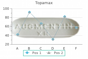
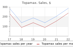
Buy topamax 200 mg without prescription. MS program on Nutrition Self Aspects & New Descriptions of Symptoms and Symptom Management.
Syndromes
- Allergic reaction to contrast dye
- Be able to act witty and charming
- Endoscopy of the abdomen
- Exercise regularly.
- When bending backward or walking more than a few yards
- Urinalysis and a urine culture (clean catch)
- Chest tightness
The proper primary bronchus and bronchus intermedius are divided and the right upper lobe is eliminated medications xerostomia purchase topamax 100 mg on line. There are two bronchial resection margins treatment 247 safe 100 mg topamax, proximal (main stem) and distal (bronchus intermedius) that should be submitted for pathological analysis treatment 100 blocked carotid artery purchase topamax line. A segmentectomy is defined as an anatomic surgical resection kerafill keratin treatment purchase topamax line, including a small segmental bronchus and artery symptoms zoning out buy generic topamax 200 mg on line. The segmental bronchus and artery must be identified on the specimen and sampled as surgical resection margins medicine zyrtec buy topamax 100 mg low cost. Simple lobectomy is the commonest surgical procedure performed for stage I lung cancer and in some particular cases of non-neoplastic disease. In this occasion, there are two bronchial margins that should be pathologically evaluated. Pneumonectomies are performed on a choose group of patient with malignant disease, as properly as for some particular cases of non-neoplastic illness. Explants from lung transplant recipients, in addition to autopsy specimens, are also included on this class. It is greatest to have these bigger specimens brought directly to the laboratory in a contemporary state throughout working hours. Specimens could be briefly stored in a dedicated refrigerator through the night and processed the following morning. Processing of those large specimens for non-neoplastic illness or suspected lung cancer will be discussed right here. Unless bronchial or vascular anomalies need to be evaluated in an unfixed specimen, formalin inflation through the bronchus or trachea is recommended. This can be completed via gravity drainage, which makes use of a plastic tube attached to a formalin tank 2:three ft above the specimen, or with a large formalin-filled syringe. The plastic tube or syringe is wedged into the airway and the specimen is filled with formalin till maximally expanded. In a centrally obstructing lesion, it might be essential to use a syringe and needle for localized formalin distension of the distal parenchyma or previously incised areas. One should at all times consider the need for special research whereas the specimen continues to be contemporary. If fresh tissue is required for microbiology or other functions, small pieces of peripheral lung may be obtained, without considerably affecting formalin distension. Extra trimming could additionally be required to completely open the vessels and smaller airways. This slab part of lung from a patient with cystic fibrosis vividly demonstrates bronchiectasis and purulent mucous plugging. This section of decrease lobe, cut utilizing the bronchial probe technique and trimmed, demonstrates bronchiectasis. The main stem bronchus is trimmed after which the hilar floor of the lung is laid down on the slicing board. The lung is serially sliced in a longtime airplane with as thin and even slices as potential. The anteriorto-posterior, apical-to-inferior dimension demonstrates a maximum amount of lung and is superb for appreciating regional variations (upper vs. Other particular orientations, similar to sagittal slicing, are used to demonstrate and evaluate emphysema and different diffuse pulmonary ailments. Although some pathologists may have a strong private choice by habit or coaching for "breadloafing", there are some compelling reasons to make the most of one of the two above-mentioned techniques. These techniques allow for the rational orientation of the lung and facilitate extra accurate gross descriptions. These kinds of orientation help to keep away from common trainee problems, such as complicated a post-obstructive pneumonia with the tumor mass, lacking airway lesions, and confusing lymph node metastases with primary tumors. This orientation additionally allows for a higher appreciation of the illness geography, as with centrilobular emphysema and pulmonary fibrosis. Lungs fastened and reduce in these manners are additionally wonderful for instructional demonstrations or pictures. As with other kinds of organs, the picture is finest when taken in an anatomically right orientation. If the specimen is being photographed in a recent state, extra blood should be blotted away. A paper towel soaked in alcohol could be briefly applied to the surface so as to reduce the sheen from a fresh specimen and may be equally utilized to a formalinfixed specimen to brighten the colours. There are alternate options to formalin fixation or sectioning which could be specifically indicated, notably for analysis. Some of those techniques have been summarized in earlier editions of this e-book, as nicely as in other common reference publications. Evaluation of the large specimen in recognized or suspected cases of major lung carcinoma the prognosis of malignancy is now typically confirmed prior to resection. The specimen was obtained from the explanted lung of a transplant recipient with extreme continual obstructive pulmonary disease. The tissue was inflated, frozen in liquid nitrogen vapor, fastened in 1% glutaraldehyde resolution, post-fixed in 1% osmium tetroxide, and critical-point dried. It is prudent to know as a lot about the radiological abnormalities as possible, previous to gross evaluation. Chapter 25 addresses the lung cancer staging system but there are a number of elementary points that ought to be made within the context of specimen handling. It is particularly important that pathologists and their trainees understand the importance of accurate tumor measurement and its potential influence on adjuvant therapeutic choices and overall prognosis. T2 tumors are subclassified into T2a (> 3 and 5 cm), T2b (> 5 and 7 cm) and T3 (> 7 cm). In addition, it was noted that not all lung tumors confirmed shrinkage after formalin fixation or even a constant fixation rate. The pathological criteria and use of elastic stains to consider visceral pleural invasion was lately reviewed on behalf of the International Staging Committee. Such a practice ensures that one can discern the true visceral pleural floor from subpleural fibrosis or incompletely minimize histological sections. In many peripheral lung tumors, visceral pleural puckering is obvious and ink must be applied generously to this space. If a frozen part is requested (or the surgeon feels it essential to cut into the specimen for some other reason), the lesion must be sampled in an area away from essentially the most clearly affected visceral pleura to avoid disruption artifacts. Pre-ordering an elastin stain on sections most suspicious for visceral pleural invasion will enhance turnaround time and end in extra correct staging. Although not a formal a half of the lung cancer staging system, thoracic pathology specialists additionally suggest that each one recognized or potential adenocarcinomas 2 cm or much less be submitted for pathological analysis of their entirety. This allows full morphological analysis and identification of tumor subtypes, which can carry therapeutic and prognostic significance. N2 and N3 nodes are mediastinal lymph nodes that should be separately recognized and designated by the surgeon (see Chapter 25). N1 nodes embody peribronchial and hilar lymph nodes connected to lobectomy specimens. These N1 nodes are extra easily identified in a specimen with a peripheral lung tumor however can be quite difficult to determine in a more central tumor. Often, significantly with an inexperienced trainee, a metastasis to contiguous N1 lymph nodes is ignored macroscopically. The carbon pigment current in most of these nodes further facilitates lymph node recognition. This type of sectioning also highlights the secondary pathologies of post-obstructive pneumonia, bronchiectasis, and 48 Chapter 2: Lung specimen dealing with and practical issues consultant samples. Many surgeons are reluctant to biopsy the lung in such circumstances, if it can be recognized amidst the dense fibrosis, for fear of a chronic air leak. However, deeper "interface" biopsies of the chest wall may be taken with minimal morbidity. Smaller biopsies ought to be submitted in toto however sampling of larger decortication or pleurectomy specimens requires data of the clinical history and an knowledgeable gross examination. Larger pleural specimens should be carefully examined for peripheral adherent lung and skeletal muscle. The variety of sections ought to correlate with the index of suspicion for malignancy. It is straightforward to recognize both the primary tumor mass and the gross involvement of lymph nodes when the specimen is reduce alongside the airway. The distance from the bronchial or medial resection margin can be more simply famous. Depending on the indication, larger specimens could also be despatched to the pathology laboratory for processing. Receipt of those larger samples is often seen with aid by each common and expert pathologists. They each perceive that with pleural biopsies, larger biopsies normally lead to a higher level of diagnostic confidence. This is particularly true when a thoughtful surgeon, an attentive pulmonologist, and an astute radiologist contribute to the analysis. Benign and malignant pleural ailments are covered in depth in Chapter 36 and subsequently only some primary principles of processing are warranted. It is preferable to include no much less than one wedge biopsy of the lung that can assist to outline the visceral pleural pathology in relationship to the underlying lung. In addition multiple biopsies from the parietal pleura, chest wall, mediastinum, and diaphragm must be taken. When the pleural space is partially or utterly obliterated, the surgeon will face higher technical difficulty in assessing the pleural illness (diffuse pleural fibrosis versus malignancy) and acquiring the appropriate use of ancillary research must be grounded in a well-formulated differential prognosis and correlated with morphology. The clinician and the radiologist contribute greatly by providing a whole and accurate history, which helps the diagnostic work-up and avoids unnecessary checks. In some areas, corresponding to interstitial pulmonary fibrosis, a combined diagnostic strategy in a joint assembly is the only rational approach to achieve the right diagnosis. A basic overview is provided here; extra particular indications for these ancillary tests are covered in subsequent chapters. Despite a great deal of technological innovation and enthusiasm for molecular testing, histochemical stains remain a mainstay within the analysis of neoplastic and non-neoplastic lung illness. Histochemical stains have the nice virtues of speedy turnaround time, low price, technical simplicity, and applicability to normal cytology and histology samples. Histochemical stains may be divided into these routinely performed to detect microorganisms (Grocott methenamine silver, Ziehl-Neelsen, Gram), these staining matrix substances (Masson trichrome, Movat pentachrome, elastic van Giesen, and Congo red), people who demonstrate intracellular mucin (mucicarmine, periodic acid-Schiff), and people detecting different substances, corresponding to iron or calcium. Immunohistochemistry has also been utilized to the diagnosis of mycobacterial, fungal, viral, and bacterial infections. Immunohistochemistry in pulmonary pathology has been prolonged into prognostic markers and therapeutic response predictors. The technique is based on a main antigen-antibody reaction and a secondary antibody-enzyme complicated, which interacts with a chromogen for a microscopically seen shade response. Immunohistochemistry may be performed on either frozen or forty nine Chapter 2: Lung specimen dealing with and practical concerns formalin-fixed tissue, relying on the antibody. It is good apply to not use a tissue block that contains previously frozen material for immunohistochemistry, if other tissue blocks are available. Unfortunately, there are only a few antibodies which strategy 100% sensitivity and specificity. This requires scientific interplay and morphological experience in generating a differential prognosis, in addition to important assessment of the immunohistochemical outcomes with acceptable controls (see Chapter 26). Immunohistochemistry is usually used within the work-up of lung and pleural tumors, either to higher characterize the first or to exclude metastatic disease. It is important to understand the staining patterns of the normal lung and pleura, notably in a small or distorted specimen. For example, cytokeratin antibodies will stain benign bronchial and alveolar epithelia, in addition to reactive mesothelial cells, which may be entrapped inside tumors and lead to a false positive interpretation. The histopathologist should pay consideration to the staining for a specific monoclonal antibody in tumors within the differential prognosis. Thus calretinin, whereas positive in some epithelioid mesotheliomas, might stain the nuclei of some sarcomatoid carcinomas (see Chapter 36). In particular person cases, immunohistochemical evaluation remains an exercise in possibilities and may not be enough for certain scientific circumstances. New markers are sometimes launched with initial printed stories of excessive sensitivities and specificities. After extra studies and incorporation into day by day practice, extra exceptions seem. The diagnosis of epithelioid mesothelioma is satisfactorily confirmed with an immunohistochemical panel in most cases. Electron microscopy, if out there, can be reserved for the occasional cases during which the immunohistochemical profile is equivocal. Prompt fixation in a recommended fixative (glutaraldehyde or methanol-free formaldehyde) is most popular, although commercial formalin could additionally be used if the fixation is rapid. A new sharp blade should be used and minimize with a to-and-fro motion, quite than by exerting downward pressure. Other selected makes use of in non-neoplastic disease embody the demonstration of electron-dense deposits. New molecular techniques for the analysis of neoplastic and infectious diseases have been introduced into the pathology laboratory, although the degree to which these techniques have been integrated into clinical apply varies worldwide. The potential purposes for immunohistochemistry and in situ hybridization have been similarly expanded. Fewer studies tackle the cost-benefit evaluation related to new assay validation and implementation into the medical laboratory. Another problem that have to be addressed inside the medical laboratory and in collaboration with scientific colleagues is what to test. The new molecular technologies can be applied to a bewildering array of different recent biological samples, including tissue, aspirates, effusion fluid, and bronchial specimens.