

Charles Williams Flexner, M.D.
- Director, AIDS Clinical Trials Unit
- Deputy Director, Institute for Clinical and Translational Research
- Professor of Medicine

https://www.hopkinsmedicine.org/profiles/results/directory/profile/0001766/charles-flexner

Functional endoscopic-approach to inflammatory sinus disease: current perspectives and approach modifications allergyworx discount 5 mg clarinex otc. Computed tomographic dacryocystography utilizing topical contrast media for lacrimal system visualization allergy medicine menstruation discount 5mg clarinex visa. Determination of the "incidental" Lund score for the staging of chronic rhinosinusitis allergy medicine name brand quality 5 mg clarinex. A comparison of symptom scores and radiographic staging techniques in chronic rhinosinusitis allergy treatment yorba linda ca purchase clarinex 5 mg free shipping. Global Osteitis Scoring Scale and continual rhinosinusitis: a marker of revision surgery allergy medicine long-term effects purchase clarinex online from canada. Diagnostic and staging accuracy of magnetic resonance imaging for the assessment of sinonasal disease allergy medicine restless leg syndrome purchase 5 mg clarinex with visa. Chronic inflammatory sinonasal illnesses together with fungal infections: the function of imaging. The significance of sinonasal radiodensities: ossification, calcification, or residual bone Analyses of distant metastases in squamous cell carcinoma of the pinnacle and neck and lesions above the clavicle 41. An assessment of sinonasal anatomic variants doubtlessly associated with recurrent acute rhinosinusitis. Anatomy of the frontal recess and frontal sinus with three-dimensional reconstruction. Endoscopic Sinus Surgery: Anatomy, Three-Dimensional Reconstruction, and Surgical Technique. Rhinology 2008;46(2):116�120 a hundred thirty eight Clinical Examination and Differential Diagnosis in Rhinology Joaquim Mullol, Franklin Mari�o-S�nchez, Isam Alobid, and Christos Georgalas Summary. Other sinonasal issues essential within the differential prognosis are nonallergic rhinitis, acute and chronic infections, structural abnormalities, vasculitides, and granulomatous ailments, in addition to benign and malignant tumors (Table 8. In this chapter, we summarize the primary diagnostic instruments which would possibly be used within the differential diagnosis of nasal and sinonasal disease, including clinical nasal symptoms (their definition and classification, etiology, and severity), nasal signs and scientific nasal examination, imaging, high quality of life, and evaluation of nasal patency and the loss of smell. In addition, we propose algorithms for multidisciplinary session and referral. Following a regular routine and systematizing the above, as seen in the following case examine, may help to avoid omissions. Case Study Patient: John Doe, a 24-year-old male instructor Presenting criticism: watery rhinorrhea History of the presenting complaint: unilateral, fixed; also occurring throughout sleep. No associated nasal obstruction, sneezing, epistaxis, or complications Past medical history: briefly hospitalized 6 months ago for concussion. There are questionnaires obtainable that can assist in the prognosis of allergic rhinitis. In different sinonasal illnesses, distinguished symptoms could additionally be purulent rhinorrhea and nasal crusting, bleeding, or obstruction (vasculitis and tumors); facial hypoesthesia (tumors); and malocclusion (facial fractures and tumors). Composites of total nasal, total nonnasal, and global scores additionally could be obtained. Rhinorrhea Nasal discharge is a typical symptom of the common chilly, allergic and nonallergic rhinitis, and rhinosinusitis (either acute or chronic). Also, it is very important decide the localization of the discharge (uni- or bilateral), the time of onset, in addition to precipitating and mitigating elements. An acute watery bilateral discharge is normally related to a typical cold or, if continual, allergic or nonallergic rhinitis. Unilateral watery rhinorrhea, particularly when elicited by bending and related to salty taste, is highly suggestive of. Note the in depth destruction of the nasal mucosa with areas of granulation and bleeding. A choanal atresia in the first days of life can interfere with breastfeeding and cause respiratory misery. In older children (often secondary to adenoid hypertrophy), nasal obstruction may be associated with ear air flow issues, affecting listening to and speech growth, and also may (together with oropharyngeal obstruction) cause. In older kids and adults, it may impression very significantly upon their quality of life by inflicting serious discomfort and by altering sleep patterns, together with the senses of scent and style. In historical past taking, it is important to decide whether nasal obstruction has been current for a very long time or if it is of latest onset, as an example, after a nasal trauma, which would strongly counsel a septal deviation after a nose fracture. Long-standing, fixed nasal obstruction (uni- or bilateral) with only a few other coexisting nasal symptoms can be the outcome of structural abnormalities, such as septal deviation, however extra often is brought on by mucosal swelling/hypertrophia of the turbinates caused by rhinitis/rhinosinusitis. Progressive over months or a quantity of years, fixed, unilateral nasal obstruction may be associated with nasal tumors, whereas intermittent, reversible nasal obstruction with distinct precipitating elements suggests an inflammatory etiology. The examiner should also elicit whether or not the obstruction is alternating between the sides (physiologic nasal cycle), unilateral or bilateral, and constant or intermittent. Fluctuation usually suggests an inflammatory mucosal situation quite than a mechanical obstruction. Nasal obstruction brought on by allergic rhinitis or rhinosinusitis is common bilateral and alternating, exacerbating the physiologic nasal cycle. If a unilateral mass is found, the etiology must always be clarified with imaging and (unless a vascular tumor is suspected) a biopsy. A bilateral nasal obstruction presents as a medical emergency in the neonate, whereas a unilateral choanal atresia can current later in life, and even throughout maturity. In a younger boy or male adolescent, unilateral nasal obstruction accompanied by nosebleeds is often attributable to a nasopharyngeal angiofibroma. There are some validated questionnaires obtainable to consider the subjective sensation of decreased airflow and its impression on high quality of life. In maxillary sinusitis, the pain is extra intense over the maxillary sinuses and the adjacent midface and zygomatic area, however it might additionally radiate to the forehead. In dentogenic maxillary sinusitis, the dental pain can be extra intense and obscure the sinogenic ache. The ache is often much less intense than that associated with maxillary or frontal sinusitis. In sphenoid sinusitis, pressure could also be situated on the middle of the cranium, vertex, or occipital region. Typical features that recommend that a headache is sinogenic embrace its affiliation with nasal symptoms, its exacerbation with higher respiratory tract infections, and its response to medical or surgical therapy of rhinosinusitis. Nonsinus headaches incessantly are characterized by the same signs, thus making it troublesome to determine if the headache is really attributable to a sinus problem. However, some symptom features may help the examiner make the differential diagnosis. Tension headaches will happen in a band shape that goes from the forehead to the neck and is normally worsened or precipitated by psychological stress. Migraine headache may be very intense, typically pulsating and (in the case of classic migraine) preceded by aura, situated behind and around the eyes, affecting one facet of the head, associated with nausea, and often lasting hours or days. Cluster headache is characterized by very severe unilateral orbital, supraorbital, and/or temporal pain assaults that happen collectively in bouts. Hyposmia/Anosmia Symptoms of olfactory dysfunction appear often in rhinologic pathologies. The sense of smell reveals a clear impairment in higher airway inflammatory diseases, ranging from moderate in allergic rhinitis to severe in nasal polyposis. Kallman syndrome is a rare explanation for congenital Clinical Diagnosis 137 anosmia; it should be included within the differential diagnosis of each patient presenting with anosmia since start. Eye Symptoms Eye symptoms may level to allergic rhinoconjunctivitis (watery, itchy eyes), nasolacrimal duct obstruction (epiphora), orbital ground blowout or silent sinus syndrome (enophthalmos with or without diplopia), sphenoid, maxillary or ethmoid tumors or mucoceles, intraorbital hematoma or abscess, or Graves disease (unilateral or bilateral exophthalmos, lowered eye mobility, and diplopia). Orbital fractures that embody the infraorbital rim usually present with hypoesthesia in the space of V2 distribution. Sneezing Upper respiratory infections and allergic rhinitis are the commonest and attribute causes of sneezing. Sneezing associated with nasal itching is very suggestive of an allergic etiology, particularly if there are coexisting eye symptoms (itching and epiphora). In many cases, nasal symptoms precede by a few years the onset of the systemic features of the illness (see Chapter 35). Classically, the severity of sinonasal illnesses (mainly for rhinitis and rhinosinusitis) has been assessed primarily based on symptom intensity. Examination and Complementary Diagnostic Tools Nasal Examination Etiology Allergy is recognized by the identification of the clinically related allergen, pores and skin prick test11 or serum-specific IgE, Although nasal examination by its nature is subjective, validated scales can be utilized as an goal measurement in assessing the colour of the nasal mucosa and the presence of septal deviation or perforation, turbinate and adenoid hypertrophy, international bodies, epistaxis, secretions from polyps within the center meatus, and benign and malignant tumors. Anterior rhinoscopy may be performed, but nasal endoscopy, either inflexible or versatile, is taken into account the gold normal. External Inspection the clinical examination should all the time start with a visual inspection of the nose, which may reveal important information. A horizontal sulcus within the pyramidal dorsum could indicate constant rhinorrhea or itching in a affected person with allergic rhinitis. It is associated with systemic illnesses corresponding to Wegener granulomatosis, congenital syphilis, polychondritis, and large septal perforations secondary to cocaine abuse or nasal surgical procedure. Advanced sinus tumors can infiltrate the subcutaneous tissue and the pores and skin and present as an exophytic ulcerated pores and skin lesion. The hazard triangle of the face, also referred to as Filatov triangle (the area from the corner of the mouth to the glabella, together with the nostril and maxilla), is a risk region for dermatologic infection, corresponding to cellulitis or furuncles, which present with circumscribed redness and swelling 140 8 Clinical Examination and Differential Diagnosis in Rhinology I Basic Science and Patient Assessment * a b *. External inspection of the nose might reveal anatomical variations and main deformities of the external nasal shape, an apparent deviation of the nasal septum, collapse of the nostrils throughout inspiration, or nasal vestibular stenosis in sufferers with cleft palate. A rough indication of nasal airflow may be obtained from the nasal exhalation pattern on a chilly metal surface, corresponding to a tongue depressor or a mirror. The Cottle maneuver, when carried out delicately, can assess the presence of nasal valve collapse. Palpation Palpation is essential in patients with recent facial trauma for detecting step-offs in a fracture line of the midfacial bones and for assessing mobility, crackling, or discontinuity within the nasal bones, which may recommend a pyramidal fracture. In sufferers with facial neuralgias, the examiner should pay attention to tenderness over the supraorbital, infraorbital, or mental foramina. Small swellings over the nerve trunk after it emerges from the foramen, in addition to painful points with or without palpable changes, are sometimes found over the nerves. When inner valve collapse is suspected in a affected person with nasal obstruction, performing the Cottle check is indicated. If the affected person can breathe better with this maneuver, the issue might be within the internal nasal valve. However, when the maneuver is completed too forcefully, every patient with or without nasal valve collapse will indicate that respiration is best. When a lack of tip help is suspected, the tip elevation test could additionally be performed by pushing the nasal tip upward at a straight nasolabial angle. If the affected person exhibits enchancment in nasal respiration with the maneuver, a scarcity of cartilaginous tip help must be suspected. Findings Anterior rhinoscopy is the recommended first approach to the nasal cavity. It can reveal necessary details about the anterior part of the nostril, where the majority of respiration problems are. When a mechanical problem causes nasal obstruction, normally the issue is in the anterior septum or within the anterior a half of the inferior turbinate, within the so-called nasal valve. The Kiesselbach plexus is the point at which numerous small branches of the external and internal carotid arteries meet. It is the most typical website for anterior epistaxis, and outstanding vessels on this area should be noted. The traits of the mucosa of the inferior turbinate might range from the bluish edematous mucosa seen in those with allergic rhinitis to the erythematous edematous mucosa seen in these with rhinosinusitis. Nasal Endoscopy Currently, nasal endoscopy is the gold commonplace and essentially the most priceless device within the analysis of the vast majority of rhinologic pathologies. With higher diameter, the quality of the picture is best, but the patient experiences extra discomfort. Assorted viewing angles are also obtainable: 0and 30-degree endoscopes are often most popular, while 45-degree endoscopes are used for examination of the maxillary sinus and frontal recess. An endoscope of 70, 90, or one hundred twenty degrees can be utilized to transorally evaluate the nasopharynx in youngsters. Anterior Rhinoscopy Technique A proper mild source, preferably a forehead gentle, is necessary for an accurate analysis. There are two principal methods of holding the Killian speculum for examination of the nose: � With the blades in a vertical place: this allows a wider view to the nasal fossa, however it can cause 142 8 Clinical Examination and Differential Diagnosis in Rhinology I Basic Science and Patient Assessment Flexible endoscopes can be used to examine the nostril, nasopharynx, hypopharynx, and larynx in one sitting. Their relative drawback in comparison with rigid endoscopes is a lower picture decision, though present expertise has made them a wonderful software offering a more than good image quality. Another disadvantage is that repeated use can break inside fibers and worsen the image quality. Often deviation of the septum and/or a slender nasal cavity allows this to be accomplished adequately on only one side of the nostril. For a detailed description of the three-pass approach, (see Video 10, Normal Three-pass Endoscopy). Direct visualization of the paranasal sinuses is very difficult when sinus surgical procedure has not been performed. However, the primary strategy must be carried out with out using vasoconstrictor substances to consider the precise dimension of the turbinates and the characteristics of nasal mucosa and secretions. First cross: Nasal endoscopy begins with the light introduction of the scope via the nostril, in an anterior to posterior direction, parallel to the nasal floor, below the inferior turbinate, attempting not to contact the septum and advancing by way of the choanae to the nasopharynx. With the endoscopy in the nasal valve, nasal valve operate with normal and extra forceful respiration could be examined. In the nasopharynx, the examiner ought to inspect the torus tubarius, eustachian tube orifice, posterior pharyngeal wall, and roof of the nasopharynx. The velopharyngeal function may be evaluated by asking the patient to repeat the letter p because the palate ascends and contacts the posterior pharyngeal wall. The presence of secretions in the nasopharynx, in addition to the general status of the mucosa, have to be famous. After analyzing the cavum, the scope have to be slowly pulled backward and barely upward following the edge of the middle turbinate to see the pure ostium of the sphenoid sinus (7 mm from the superior border of choanae, behind the superior turbinate), middle and superior turbinates, and ethmoid cells.
In the absence of urinary retention or neurological options allergy medicine 6 month old buy clarinex, assume detrusor overactivity because the doubtless prognosis allergy treatment under the tongue purchase clarinex now. Finally allergy virus buy clarinex 5 mg lowest price, decide the level of distress caused by signs and the general influence on high quality of life Consider referral for complete geriatric evaluation allergy testing your baby purchase clarinex in india. Bleeding may be due to quinolone allergy symptoms best order clarinex miscarriage or ectopic being pregnant allergy to eggs buy cheap clarinex 5 mg, or could have a benign explanation in the presence of an ongoing intrauterine pregnancy. Miscarriages may be threatened (the foetus continues to be viable), partial (non viable products of conception remain in the uterus) or complete (products of conception have been passed). Miscarriages usually current with central, crampy, menstruallike decrease belly ache In ectopic being pregnant, the pain is unilateral and fixed since there could also be bleeding into the peritoneal cavity inflicting peritoneal irritation. The goal definition is blood loss higher than 80 mL in a cycle, however in apply prognosis relies on ladies subjectively reporting utilizing more tampons/pads than traditional, using pads and tampons together or soaking via protection onto clothing. Structural causes embody fibroids (leiomyomas) (20�30%), endometrial and cervical polyps (5�10%), adenomyosis (5%) and malignancy or hyperplasia. These may be categorized broadly in accordance with the reproductive age of the patient, their pregnancy standing, the period of signs and any related features. Examine the affected person fastidiously to determine rectal, urethral, vaginal and cervical sources of blood; if needed, place a urinary catheter to differentiate between vaginal bleeding and haematuria. In this situation, blood loss may still be significant however remains contained within the uterine cavity. The sample of bleeding may be associated to contact such as postcoitus, or intermenstrual bleeding with no clear precipitants. Causes of contact bleeding are usually cervical, corresponding to cervical ectropion, cervical most cancers, cervical polyps or infections (typically chlamydia but also gonorrhoea). There is a continuum between intermenstrual and irregular menstrual bleeding that can be difficult to differentiate. Oral contraception and intrauterine system ks fre Intermenstrual bleeding contraception might trigger abnormal menstrual bleeding. Uncommonly, low-volume mid-cycle bleeding could also be due to ovulation (when the Graafian follicle ruptures and hormonal stability becomes predominantly progesterone-based, resulting in a shed of blood from the endometrium). Other malignant causes include cervical, vaginal, vulval and, rarely, ovarian most cancers. Atrophic vaginitis with contact bleeding because of postmenopausal dryness, pessary devices or intercourse is a analysis of exclusion. Yes co m Vaginal bleeding Step-by-step assessment 291 fre Clinical tool Examination of the feminine genitalia the vaginal examination of females with an intact hymen must be avoided, notably as the knowledge requ purple can typically be obtained by digital examination of the ectum. Its intimate nature raises medicolegal concerns necessitating both informed consent and the presence of a chaperone all through the examination. When performing bimanual and speculum examination, assess the quantity of handed blood, verify for adnexal tenderness (ectopic pregnancy) and establish products of conception on the cervix that require elimination. Remember that younger sufferers, particularly pregnant women, could keep comparatively normal physiological parameters until shock is profound. Identify sources of lower reproductive tract (endocervix vagina, vulva) bleeding through careful examination and biopsy any seen lesions. Biopsy all visible cervical lesions even when the cytological smear is seemingly regular. Take vaginal/cervical swabs if the history raises suspicion of sexually transmitted an infection. Enquire rigorously about associated postcoital or intermenstrual bleeding � if present assess as per step 5. Arrange cervical smear cytology except lately carried out and biopsy any visible cervical lesion even if the cytological smear is outwardly regular. Establish the degree of misery caused by bleeding and the impression on quality of life as this may influence treatment choices. Surgical options embrace endometrial ablation, myomectomy (removal of fibroids), or, if all different avenues are unsuccessful and the affected person wishes for no more children, hysterectomy. Intermenstrual bleeding unrelated to intercourse/contact is more likely to have an endometrial or systemic trigger. Enquire about contraception use and, in particular, any recent modifications: the mixed oral contraceptive pill typically causes breakthrough bleeding (small quantity recognizing between the monthly bleed when omitting the tablets for 7 days). Progesteronebased contraceptives (depot preparations similar to Nexplanon, hormonal coil corresponding to Mirena) typically result in an irregular pattern of bleeding. Consider screening for coagulopathy if there are suggestive options (see step 4). Refer any affected person with persistent unexplained intermenstrual bleeding for gynaecology evaluation and further investigation. Patients with hypercalcaemia might experience weight loss because of underlying malignant disease or the direct results of Ca2+. In peptic ulcer illness, mesenteric ischaemia and continual pancreatitis, the pain related to consuming might result in weight loss through avoidance of food. Limb weak spot and impaired functional talents can present a significant barrier to food preparation and consumption. Multifactorial weight loss can also be frequent in dementia, particularly in superior stages. The prevalence of melancholy in older adults (>65 years old) is estimated at around 35%, and better nonetheless in these living in institutional settings. Addressing the underlying depressive illness can lead to improved oral intake and weight gain. Lesser degrees of weight loss may also signify underlying pathology, significantly in the frail aged. Establish how a lot weight has been lost, and over what time frame; whether or not there has been a change in garments dimension; and whether or not there was a corresponding change in urge for food. Weight loss can be one of several non-specific presenting symptoms in continual adrenal insufficiency (Box 25. Empagliflozin oo m bo ok s Direct weight loss impact sf re Other Explore attainable social causes for weight loss such as lack of cash to purchase meals social isolation and/or chronic alcohol extra Poor dentition or ill-fitting dentures can also contribute to undernutrition. Refer, initially, to the related chapter if one of many following signs is current: Breast lump (Ch. In the absence of ketoacidosis, check with your native diabetes service for remedy initiation. Remember that a mild, transient enhance in blood glucose stage may occur in any extreme acute illness (stress hyperglycaemia). In sufferers with identified diabetes, use plasma HbA1c to gauge the severity of recent hyperglycaemia; contemplate hyperglycaemia as a contributor to weight loss if management is suboptimal (especially in kind 1 diabetes) but continue to search for alternative causes. Look for clues in the history, physical examination and routine tests that may assist to target further investigation. Other causes, notably if hypercalcaemia is gentle, embrace sarcoidosis, thiazide diuretics and vitamin D analogues. Bear in mind that localized prostate most cancers is unlikely to account for important weight loss so proceed to seek various causes unless there are features of advanced illness. Lymphadenopathy, either native or generalized, could signify infection, irritation. In sufferers with reduced dietary intake ask about signs similar to dry mouth, drowsiness, altered taste sensation or nausea and search for potential drug culprits (Box 34. If you believe you studied a drug trigger, reassess weight and signs after trial discontinuation (if appropriate). If you think despair as the cause of weight reduction reassess weight and different symptoms after a period of remedy. Where potential search collateral historical past from associates or household (who might have raised the issues re: weight loss). Explore associated components which might be contributing to weight loss corresponding to urge for food suppression, substitute of meals with alcohol intake, lack of money to purchase food or co-existent mood disorders. Explore possible contributing factors to inadequate food intake including insufficient dentition, swallowing issues, lack of motivation (or money), impaired mobility (unable to get to shop) or refined cognitive impairment. Wherever possible, get hold of a collateral history from pals, relations or carers to evaluate the contribution from cognitive impairment to weight reduction. Examine carefully and repeatedly for murmurs and peripheral stigmata of endocarditis. Be delicate and tactful but persistent in questioning and refer to specialist companies if important concern. These targets ought to, in fact, be individualized in consultation with the particular person with diabetes. These might be extra outstanding, with a shorter history, in kind 1 diabetes, as a end result of absolute insulin deficiency. In type 2 diabetes, there may be little in the way in which of signs, or they could go unnoticed due to their insidious onset. In symptomatic sufferers, one laboratory measure is required to confirm hyperglycaemia. Severe hypoglycaemia is defined by the requirement for exterior assistance to restore regular blood glucose (whether relations or emergency services). It could be sophisticated by myocardial infarction, stroke or peripheral arterial thrombosis In addition, overly speedy correction of metabolic abnormalities can lead to complications such as seizures, cerebral oedema and central pontine myelinolysis. It is, due to this fact, vital to right abnormalities gradually and to carefully monitor the plasma osmolality ([2 � Na] + urea + glucose) throughout therapy. Severity may be graded as mild/moderate/severe depending on the degree of biochemical abnormality and presence of altered mental status (Table 34. Hyperglycemic crises in grownup sufferers with diabetes: a consensus assertion from the American Diabetes Association Diabetes Care. The fundamental ideas of heredity and, as a consequence, genes could be traced back to 1865 and the research of Gregor Mendel � discussed by Orel (1995). From the results of his breeding experiments with peas, Mendel concluded that every pea plant possessed two alleles for every gene, however only displayed a single phenotype. Perhaps probably the most exceptional achievement of Mendel was his capacity to correctly establish a fancy phenomenon with no knowledge of the molecular processes concerned in the formation of that phenomenon. Hereditary transmission via sperm and egg grew to become recognized about the same time and Ernst Haeckel, noting that sperm consists largely of nuclear material, postulated that the nucleus was responsible for heredity. Both proteins and nucleic acid have been thought of as doubtless candidates for the function of the genetic materials. Firstly, proteins are plentiful in cells; although the quantity of an individual protein varies significantly from one cell type to one other, the overall protein content material of most cells accounts for over 50% of the dry weight. Secondly, nucleic acids appeared to be too easy to convey the advanced info presumed to be required to convey the characteristics of heredity. He separated nuclei from the cytoplasm of cells, after which isolated an acidic substance from these nuclei that he called nuclein. Miescher confirmed that nuclein contained massive amounts of phosphorus and no sulphur, characteristics that differentiated it from proteins. In what proved to be a exceptional insight, he advised that `if one needs to assume that a single substance. They proposed a very simple four-nucleotide unit that was repeated many times to kind long nucleic acid molecules. The tetranucleotide model for nucleic acid construction proposed by Levene and Simms in 1926. In 1928, Frederick Griffith performed experiments using a quantity of totally different strains of the bacterium Streptococcus pneumoniae (Griffith, 1928). Some of the strains used had been termed virulent, meaning that they triggered pneumonia in each people and mice. Virulent and avirulent strains are morphologically distinct in that the virulent strains have a polysaccharide capsule surrounding the bacterium and kind easy, shiny-surfaced colonies when grown on agar plates. Avirulent micro organism lack the capsule and produce tough colonies on the same plates. Griffith knew that solely dwelling virulent micro organism would produce pneumonia when injected into mice. Neither cell kind triggered dying in mice when they have been injected alone, however all mice receiving the mixed injections died. The analysis of blood of the dead mice revealed numerous residing clean micro organism when grown on agar plates. Griffith concluded that the heat-killed easy micro organism were by some means answerable for converting the stay avirulent rough bacteria into virulent clean ones. They began by culturing large portions of easy Streptococcus pneumoniae cells. Following homogenization and a variety of other extractions with detergent, they obtained an extract that, when examined by co-injection with live rough bacteria, nonetheless contained the remodeling principle. Protein was removed from the extract by several chloroform extractions and polysaccharides were enzymatically digested and removed. Finally, precipitation of the resultant fraction with ethanol yielded a fibrous mass that still retained the power to induce transformation of the rough avirulent cells. Griffith famous that injecting reside clean micro organism into mice led to the formation of pneumonia and ultimately to the death of the mouse. The phage was recognized to be adsorbed to the floor of the micro organism, after which there was a latent interval of roughly ten minutes earlier than infectious virus particles started to be made, finally resulting in host cell lysis and phage release. During the course of an infection, the bacteriophage adheres to the surface of the Escherichia coli cell. The Hershey�Chase blender experiment to show that nucleic acid was the genetic material. Hershey and Chase grew T2 bacteriophages on micro organism whose media contained either 32 P (to label the phosphorus of nucleic acid) or 35 S (to label the sulphur of proteins � the aspect chains of the amino acids methionine and cysteine both contain sulphur). They used their radio-labelled bacteriophages to infect a new tradition of unlabelled bacteria. After a short incubation, the micro organism have been harvested by centrifugation and put into a blender to shear the micro organism away from the phage particles connected to their floor. These labelled phages can be isolated from the medium of infected cultures and used to infect other unlabelled micro organism.
The bone of the anterior skull base between the two orbits is then eliminated allergy shots mechanism buy clarinex with a mastercard, thus creating a large surgical corridor allergy medicine nausea purchase clarinex 5 mg on-line, which may be extended laterally between the two medial orbital partitions allergy symptoms of beer 5mg clarinex fast delivery, and anteroposteriorly from the frontal recess to the sella allergy medicine herbal buy clarinex 5mg low cost. Middle Skull Base Through the endoscopic endonasal route allergy shots for fire ants order 5mg clarinex with amex, the center cranium base coincides with the superior allergy forecast euless tx buy clarinex 5mg with amex, posterior, and lateral walls of the sphenoid sinus; although a wider opening of the anterior wall of the sphenoid sinus, with the elimination of the superior and/or supreme turbinates and of the posterior ethmoid cells, is essential to achieve a greater exposure of such areas. Particular attention have to be paid to avoid accidents to the posterior ethmoidal artery. It can also be necessary to not prolong the elimination of the nasal septum and the ethmoid too anteriorly to avoid damaging the olfactory nerve and/or the lamina cribrosa. Thebony protuberances of the optic nerve and the intracavernous carotid artery, together with the lateral opticocarotid recess, also represent useful landmarks to recognize the medial opticocarotid recess; this structure represents the lateral restrict of bone removing above the sella. Again, between such bony protuberances,some epressionsareformed;thefirstoneislimd ited by the cavernous sinus apex and the V2 protuberances, whereas the second is enclosed by the protuberances of V2 and V3. Through the endoscopic endonasal strategy to the ethmoidal planum, the a part of the anterior cranium base enclosed between the medial wall of the 2 orbits has been uncovered. Thus, when the sphenoid cavity has been totally exposed and all of the septa within are eliminated, a series of protuberances and depressions turns into seen on its posteriorwall. Onbothsides,lateraltothesellarfloor,thetwo bony prominences of the intracavernous carotid arteries A. The posterior ethmoidal artery represents the anterior restrict of the planum sphenoidale opening. Note the close relationship between the posterior ethmoidal artery and the anterior wall of the sphenoid sinus. The extent of elimination of the tuberculum sella and/or of the sphenoid planum should be 1. After the bone has been removed, the dura over the sellarfloor,thetuberculumsella,andplanumsphenoidale are opened and the main suprasellar neurovascular structures become seen. The complete suprasellar region has been divided into 4 areas by two imaginary planes-one passing by way of the inferior surface of the chiasm and the mammillary our bodies and one other passing via the posterior margin of the chiasm and the dorsum sellae. In the suprachiasmatic region, as quickly as the dura is opened, the chiasmatic and the lamina terminalis cisterns appear. In the chiasmatic cistern, the anterior margin of the chiasm and the medial portion of both optic nerves are observed. The whole spectrum of anterior cerebral arteries, A1 and A2 segments of each side, the anterior speaking artery, and the recurrent Heubner arteries, together with the gyri recti of the frontal lobes, are recognized after the lamina terminalis cistern is opened. The lamina terminalis could be uncovered by 540 Rhinology enlarging the space between the anterior speaking artery and the chiasm. In the subchiasmatic area, immediately after breaching the arachnoid between the optic nerves, the pituitary stalk is encountered under the chiasm. The origin of the ophthalmic artery beneath the optic nerve can also be seen, and extra laterally and deeply, theinternalcarotidartery,itsbifurcation,andthefirstA1 phase. As a matter of reality, the superior facet of the pituitary gland and the dorsum sellae are also visible. In the retrosellar area, by passing the endoscope between the pituitary stalk and the inner carotid artery. The sellar ground has been utterly eliminated to mobilize the gland throughout dissection maneuvers. The diaphragma sellae has been eliminated and the third cranial nerve coming into the cavernous sinus comes into view. The thalami, which characterize its lateral partitions and the massa intermedia among them, the foramina of Monro anteriorly, the pineal and the suprapineal recesses, the posterior commissure, the habenular commissure, the habenular trigone, and the beginning of the aqueduct, are visualized posteriorly. The nasal mucosa is indifferent from the vomer, alongside the inferior wall of the sphenoid sinus, and bilaterally and superiorly to determine the vidian nerves that symbolize the lateral limits of the surgical corridor. The vomer and the inferior wall of the sphenoid sinus have to be eliminated fully in an anteroposterior path to allow the publicity of both the sphenoidal and rhinopharyngeal elements of the clivus. The vidian nerves, after crossing the intrapetrous carotid artery from above, attain the pterygopalatine fossa at a stage inferior to that of the intrapetrous carotid artery. For this purpose, they characterize a useful landmark during the bone removal of the inferior wall of the sphenoid sinus as a result of, by remaining medial to these nerves, the risk of injuries to the intrapetrous portion of the carotid artery is averted. Inside the sphenoid sinus cavity, the lateral limit of the sphenoid portion of the clivus is represented by the paraclival tracts of the intracavernous carotid artery, that are clearly seen. The clival bone contains essentially the most intensive collection of intercavernous venous connections across the midline. By removing extra bone as a lot as the carotid protuberances, it turns into potential to acknowledge the sixth cranial nerve. This nerve enters the cavernous sinus by passing through the basilar sinus close to the paraclival tract of the intracavernous carotid in close contact to the dorsal meningeal artery. The latter is a branch of the meningohypophyseal artery and offers arterial blood provide to the dura of the clival region. Furthermore, extending the clival bone opening downward, the anterior floor of the craniovertebral junction is reached. To provide a wider publicity of this area, nonetheless, the nasopharynx and the vomer could be completely removed as much as the exhausting palate. Removing the lower third of the clivus up to the occipital condyles enables both foramina lacera to be uncovered. The anterior third of the occipital condyles can be eliminated along with the anterior and middle third of the condyles. Access is possible by the means of endoscopic strategies through the use of a decrease trajectory, as in contrast with that essential for the sellar area. The clivus is split by the sphenoid sinus inferior wall in two portions-that is, the sphenoid (upper clivus) and the rhino-pharyngeal (lower clivus) phase. After exposure of the retrosellar area, the ground of the third ventricle has been opened on the stage of the tuber cinereum. Advancing the endoscope through this opening and beneath the massa intermedia, the posterior part of the third ventricle is visualized. After dissecting the muscular structures, the anterior arch of the atlas is eliminated and the dens is exposed. By using the microdrill, the dens can be thinned, separated from the apical and alar ligaments, dissected from the transverse ligament, after which removed. The intradural segmentofthevertebralartery,afteremergingfromthefibrous dural canal, ascends in front of the rootlets of the hypoglossal nerve to attain the anterior facet of the medulla oblongata the place it unites close to the junction of the pons and medulla with its homologue to kind the basilar artery. Below the doorway of the vertebral artery in the canal, the dentate ligament is hooked up to the pia mater between the dorsal and ventral rootlets of the spinal twine and presents a collection of triangular toothlike processes on each side that are attached at intervals to the dura mater. At the craniocervical junction, the dentate ligament is located behind the vertebral artery and the ventral rootlets of C1 and C2. B the Parasellar Region Because the endoscope provides the opportunity to clearly discover the midline of the skull base, it supplies entry to the lateral sellar compartment,54 represented mainly by the cavernous sinus. To enable a better exposure of such areas, as already beforehand described, the anterior sphenoidotomy has to be extended extra laterally and then superiorly, the supreme turbinates have to be removed, and the posterior ethmoid cells have to be opened. Alternatively, a more direct trajectory, which passes laterally to the center turbinate, might be performed to expose the lateral wall of the sphenoid sinus. Its medial wall is constituted by a fibrous trabecular construction that separates the cavernous sinus from the outer periosteal layer of the pituitary gland. The opening of such construction instantly sheds gentle on the C-shaped segment of the intracavernous carotid artery. On the opposite hand, the inferolateral trunk, with its branches to the intracavernous cranial nerves, lies laterally to the carotid artery and embedded in the lateral wall of the cavernous sinus. Lateral to the intracavernous carotid artery, the oculomotor, the abducent, and the maxillary nerves could be detected lying in a more medial aircraft in comparison with that occupied by the trochlear and the ophthalmic nerves. In actual fact, from the endoscopic standpoint, the oculomotor nerve partially covers the trochlear nerve, whereas the V1 branch of the trigeminal nerve is partially hidden behind the sixth nerve. Inferiorly, after crossing the decrease paraclival tract of the carotid artery, the trochlear nerve runs parallel to the oculomotor nerve. Nevertheless, a better anatomic description might be achieved by analyzing each area individually as separated 544 Rhinology A. Note the bone masking of the maxillary branch of the trigeminal nerve, which is properly outlined on this specimen. The lateral aspect of this area contains the fourth cranial nerve and a portion of the V1 branch of the trigeminal nerve. The ophthalmic department of the trigeminal nerve and arteries to the inferior cavernous sinus move through this area. Finally, particularly in circumstances of wellpneumatized sinuses, an inferior quadrangular space could be identified. It is restricted superiorly by V2 and inferiorly by the vidian nerve, whereas its anterior and posterior margins are represented respectively by the bony floor of the lateral wall of the sphenoid sinus from the foramen rotundum to the pterygoid canal and the intrapetrous section and caudal portion of the carotid artery. The numerous research produced in recent times on anatomic themes associated to navigating the transsphenoidal route with an endoscope bear witness to the vitality, rapid evolution, and progress of most of these contemporary surgery. Conclusion the endoscopic endonasal route is a very versatile method that provides direct visualization of the whole midline skull base extending from crista galli to the 41 Endoscopic Anatomy of the Skull Base and Parasellar Region 545 A. After the removal of the medial wall of the cavernous sinus, the third and fourth cranial nerves, operating laterally to the intracavernous carotid artery, are recognizable. After the elimination of the lateral wall of the cavernous sinus, displacing the interior carotid artery medially, are exposed the third, fourth, sixth, ophthalmic, and maxillary nerves and the inferolateral trunk, the tiny branches of which supply the cavernous phase of those nerves. The authors positively assume that laboratory cadaveric dissection remains undefeated in anatomic examine, because it presents a novel experience of a wide range of sensorial i nputs;nevertheless,ithasto be reminded that the research of surgical anatomy lately modified. It has been evolving constantly, being integrated and upgraded by the modern imaging systems, these days with the development of threedimensional computed tomography�based models. Tuberculumsellaemeningioma: a report on administration on the premise of a surgical expertise with 70 patients. Partial labyrinthectomy petrous apicectomy approach to the petroclival region: an anatomic and technical study. Surgical entry to the bottom of cranium and upper cervical backbone by extended maxillotomy. Anteriortranspetrosal-transtentorial method for sphenopetroclival meningiomas: surgical methodology and ends in 10 sufferers. The suboccipital transcondylar strategy to the clivus and cranio-cervical junction for ventrally placed pathology at and above the foramen magnum. The anterior subtemporal, medial transpetrosal approach to the higher basilar artery and ponto-mesencephalicjunction. Conservative (labyrinthpreserving) transpetrosal strategy to the clivus and petroclival region-indications, problems, outcomes and lessons discovered. Thecombinedsubtemporal-suboccipital strategy: a modified surgical access to the clivus and petrous apex. Transoral transclival removal of anteriorly placed cavernous malformations of the brainstem. Different surgical a pproaches to the sellar area: specializing in the "two nostrils fourhandstechnique. Endoscopic anatomy alongside the transnasal method to the pituitary gland and the encircling tructures. The endoscopic endonasal strategy to the lateral recess of the sphenoid sinus via the pterygopalatine fossa: comparability of endoscopic and radiological landmarks. Expanded endonasal a pproach: vidian canal as a landmark to the petrous inner c arotidartery. Endoscopic endonasal surgical procedure of the midline cranium base: anatomical study and c linicalconsiderations. Endoscopic endonasal pituitary transposition for a transdorsum sellae approach to theinterpeduncularcistern. The prolonged endoscopic endonasal strategy to the clivus and cranio-vertebral junction:anatomicalstudy. Neurosurg Rev 2007; 30(3):189�194,discussion194 41 Endoscopic Anatomy of the Skull Base and Parasellar Region fifty one. Endoscopic transnasal approach to the cavernous sinus versustranscranialroute: anatomic research. Microsurgical anatomy of the transcondylar, supracondylar, and paracondylar extensionsofthefar-lateralapproach. The e xpanded endonasal method: a totally endoscopic transnasal approachand resectionoftheodontoidprocess:technicalcase report. Endoscopic transpterygoid method to the lateral sphenoid recess: surgical approach and medical expertise. Endoscopic transnasal transpterygopalatine fossa approach to the lateral recess of the sphenoid sinus. Connections of sympathetic fibres contained in the cavernoussinus:amicroanatomicalstudy. The use of a threedimensional novel computer-based mannequin for evaluation of the endonasal endoscopic method to the midline skull base. Preliminary expertise with a brand new three-dimensional computer-based model for the study and the analysis of cranium base approaches. Otolaryngol Clin North Am 2002; 35(6):1283�1288,viii 547 forty two Pathology of the Sinonasal Region and Anterior and Central Skull Base Michael J. Berry evaluating apparent fibrous dysplasia, possible Paget illness, or involvement of the cribriform plate. Following thorough radiologic evaluation, tissue for definitive histologic prognosis is often, but not always, the following step in analysis. When accessible, tissue should be obtained transnasally, often with an endoscope. Frozen part evaluation prior to definitive surgical resection is indicated in thoughtfully chosen situations. Surgical excision might be a significant factor of the therapy of many of the pathologic entities seen at the cranium base, but not all. The role of surgery is usually limited to obtaining diagnostic tissue in cases of malignant lymphoma and selected pediatric tumors corresponding to rhabdomyosarcomas.
The maxilla bone itself articulates with eight completely different bones: frontal allergy testing types buy 5 mg clarinex overnight delivery, ethmoid allergy pollen count purchase generic clarinex pills, palatine allergy attack order clarinex once a day, nasal allergy symptoms worse in morning discount 5 mg clarinex otc, zygomatic allergy symptoms stiff joints generic 5 mg clarinex free shipping, lacrimal xyz allergy medicine order clarinex 5 mg, inferior turbinate, and vomer. The ostium of the maxillary sinus opens into the posteroinferior half of the ethmoid infundibulum in a crevice created by the uncinate process of the ethmoid. The mucus generated throughout the maxillary sinus is mobilized by the cilia in opposition to gravity, up from the sinus floor toward the maxillary sinus ostia in a stellate sample. The uncinate process of the ethmoid bone forms a significant portion of the medial antral wall throughout the center meatus. This is reported to be associated with episodes of recurring acute rhinosinusitis. Another anatomical variation is the infraorbital ethmoid cell (Haller cell), which is an ethmoid cell that pneumatizes into the roof of the maxillary sinus/floor of the orbit the sphenoid sinuses are centrally situated within the cranium base and are intimately associated to the sella turcica posteriorly, cavernous sinuses and inner carotid arteries laterally, and optic nerves superiorly. Also, the maxillary division of the trigeminal nerve and the vidian nerve neighbor the sinus. The sphenoid sinus drains through the sphenoethmoid recess, which is located within the house between the superior turbinate, septum, and cranium base. The sphenoid bone, which is situated on the most posterior portion of the nasal cavity, articulates with the ethmoid, frontal, vomer, occipital, parietal, temporal, zygomatic, and palatine bones. Pneumatization of the sphenoid sinus is classed into four classes: conchal, presellar, sellar, and postsellar, every type comprising four. The intersinus septum might lie obliquely, anchoring itself to the inner carotid artery or the optic nerve, as seen in. Separate from the intersinus septum that divides the left from the proper sphenoid are occasional incomplete septations, that are commonly inserted onto the carotid artery. Note the space lateral to the dotted red line drawn from the foramen rotundum to the canal of the vidian represents the pterygoid recess of the sphenoid sinus (orange). Note the relative position of the mastoid air cells (yellow) and petrous apex with cochlea (red circle). Occasionally, the anterior clinoid course of itself could be pneumatized, forming a recess throughout the sphenoid sinus. Again, that is of clinical significance within the administration of lateral sphenoid sinus encephaloceles associated with Sternberg canal. The posterior wall of the sphenoid sinus is part of the clivus (Latin for "slope"), which is an anatomical area comprised of the sphenoid and occipital bones extending from the foramen magnum to the posterior boundary of the sella turcica referred to as the dorsum sellae. Note the intersinus septum of the sphenoid sinus may lie obliquely, anchoring itself to the interior carotid artery or the optic nerve. A highly pneumatized posterior ethmoid cell sometimes extends posteriorly and "invades" the superior, lateral, and posterior portions of the sphenoid bone. Note the saddle-shaped melancholy of the sella turcica between the posterior planum sphenoidale and the clivus. The sphenoethmoid cell could pneumatize to a variable extent around the optic nerve, which can be manifested simply as a lateral bulge, or in extreme circumstances, appear to cross by way of the middle of the cell. It has been famous that genetic elements can also play a role, as sphenoethmoid cells appear to be more frequent in Asian sufferers. It articulates with the ethmoid, lacrimal, maxillary, nasal, parietal, sphenoid, and zygomatic bones. The drainage passageway of the frontal sinus is an hourglass-shaped area composed of three different parts. The top portion of the hourglass is the frontal infundibulum, which is the inferiormost aspect of the frontal sinus. The slender portion of the hourglass is the frontal sinus ostium that sits in the posteromedial a half of the sinus on the inferior end of the frontal infundibulum. Mucus generated within the frontal sinus circulates around the sinus in a superolateral-to-inferomedial direction earlier than draining out through the frontal recess17. Narrowing in any of those constructions or disease within the anterior ethmoid sinus can lead to frontal sinusitis. Anatomical variations are frequent within the frontal sinus and have been mentioned in nice detail elsewhere. Several ifferent classification techniques can be found for these, however none is broadly agreed upon. The commonest system is referred to as the Bent and Kuhn system, which addresses the need for a more exact definition of the frontal sinus cells separate from other kinds of anterior ethmoid cells. The agger nasi is seen because the prominence on the lateral nasal wall across from the forefront of the center turbinate. It is the intranasal representation of the ascending means of the maxilla externally. A majority of the time, this mound is pneumatized, giving rise to the agger nasi cell, which is the anteriormost ethmoid cell. Frequently, the frontal sinus is difficult to visualize endoscopically till the posterior and medial partitions of the cell are removed. When pneumatized extra posteriorly and laterally, the agger nasi cell can narrow the frontal recess and intervene with the drainage of the frontal sinus. This is a skinny crest of ethmoid bone to which the falx cerebri attaches intracranially. When pneumatized, it could drain instantly into either the frontal sinus or the ethmoid sinuses. Along the anterior table or floor of the frontal sinus on the midline, a sinus can develop called an intersinus septal cell. It is shaped by pneumatization of the frontal bone between the two frontal sinuses. The presence of the cell can slim the frontal recess on both facet, contributing to obstruction or giving rise to signs by harboring mucosal sinus illness within itself. The boundaries of the frontal recess are the posterior wall of the agger nasi anteriorly, the anterior wall of the ethmoid bulla posteriorly, the lamina papyracea laterally, and the middle turbinate medially32. The frontal recess mostly drains medial to the uncinate process and lateral to the middle turbinate into the middle meatus. The pattern of drainage and topography of this area is greatly affected by the anatomy of the anterior ethmoid sinuses. Note the place of the supraorbital ethmoid compartment (purple) to the posterior aspect of the frontal sinus area and the center turbinate (orange) connected superiorly to the lateral lamella of the cribriform plate (red circle). It frequently follows the contour of the orbital roof, pneumatizing laterally and superiorly over the orbit. The supraorbital ethmoid cell is easily mistaken for the frontal sinus correct; nevertheless, its drainage pathway is separate from the frontal recess, typically posterior and lateral to it11. The blood supply to the frontal sinus is supplied by the supraorbital and supratrochlear arteries that are derived from the ophthalmic artery. The innervation of the sinus is supplied by the supraorbital and supratrochlear nerves off the frontal nerve, which is a branch of the ophthalmic division of the trigeminal nerve. The venous drainage is to the superior ophthalmic vein, which in turn drains to the cavernous sinus. These are mucosa-lined foramina and also mark the exit points of the diploic veins of Breschet. These veins provide direct communication between frontal sinus mucosal capillaries with the dural sinuses and the marrow cavity of the frontal bone. This communication permits frontal sinusitis to turn into osteomyelitis and intracranial an infection. Note the diploic veins of Breschet provide direct communication between frontal sinus mucosal capillaries with the dural sinuses and the marrow cavity of the frontal bone. Note the dehiscence of bone overlying the left orbit (white arrows) and bony disruption of the maxillary sinuses (yellow arrows) from prior Caldwell-Luc surgical procedure with inferior turbinate resections. All of the opposite paranasal sinuses ventilate and drain by way of the clefts created throughout the ethmoid complex. The sphenoid sinuses sometimes drain via the sphenoethmoid recess, the maxillary via the ethmoid infundibulum, and the frontal sinuses via the frontal recess. At the midline, the ethmoid advanced is divided by the perpendicular plate of the ethmoid, which is steady with the cribriform plate of the ethmoid superiorly. The ethmoid sinuses are located in the upper part of the lateral nasal wall simply lateral to the medial wall of the orbit (lamina papyracea of the ethmoid). These sinuses display probably the most complex anatomy and variability of all the sinuses. The olfactory bulbs relaxation on top of the box, and the olfactory neuroepithelium lines the internal roof of the box. The ethmoid complicated articulates with 15 bones: paired frontal, sphenoid, nasal, maxillary, lacrimal, palatine, and inferior turbinate bones, along with the unpaired vomer. The boundaries of the sinus are the middle and superior turbinates medially, the lamina papyracea laterally, the perforated frontal bone superiorly, and the face of the sphenoid posteriorly32. The length of the lateral lamella of the cribriform plate determines the height of the ethmoid cavity. It is important to acknowledge that the superior extent of the anterior ethmoid roof lateral to the center turbinate could be significantly larger than the cribriform plate. The top of the ethmoid roof can be asymmetric between the left and the best sides. Keros categorised totally different configurations of the ethmoid roof with respect to the increasing length of the lateral lamella,forty five as seen in. The divisions within the ethmoid sinus are formed by a series of parallel lamellae which are obliquely oriented. The first lamella is the uncinate course of, the second is the ethmoid bulla, the third is the basal lamella of the middle turbinate, and the fourth is the lamella of the superior turbinate. As talked about beforehand, the basal lamella of the center turbinate also serves because the partition between the anterior and the posterior ethmoid sinuses. This classification system emphasizes the potential for intracranial superior and supreme meatus. The anterior ethmoid cells are generally smaller than the posterior ethmoid cells. The uncinate process is a sagittally oriented structure shaped like a hook that traverses anterosuperior to posteroinferior. The superior aspect of this structure most regularly attaches laterally to the lamina papyracea, but it could possibly entry while working medial throughout the ethmoid at its superior extent. Note that the "roof" of the ethmoid sinuses is definitely comprised of frontal bone (green), and the ethmoid complex (yellow) is highlighted by the blue circle in the central drawing. The uncinate has three layers: nasal mucosa, ethmoid bone, and infundibular mucosa. The place and orientation of the uncinate may vary depending on the anatomy of the maxillary sinus or disease process (see the dialogue of maxillary sinus above). The second lamella, the ethmoid bulla, is the biggest and essentially the most constant anterior ethmoid cell. It is situated simply posterior to the uncinate process and is predicated on the lamina papyracea. Located between the uncinate course of and the ethmoid bulla is the hiatus semilunaris, which is a crescentshaped gap between the posterior margin of the uncinate and the anterior wall of the ethmoid bulla. The boundaries of the infundibulum are the uncinate course of medially, the lamina papyracea laterally, the ethmoid bulla posteriorly, and the ascending strategy of the maxilla and lacrimal bone anteriorly8. The ostiomeatal unit refers to a group of center meatal structures rather than a specific anatomical structure. It is a practical unit fashioned by the uncinate course of, ethmoid infundibulum, anterior ethmoid cells, and ostia of the anterior ethmoid, maxillary, and frontal sinuses. An obstruction in this area could lead to illness in a number of neighboring sinuses. The posterior ethmoid sinus lies posterior to the basal lamella of the middle turbinate. It is bounded laterally by the lamina papyracea, medially by the vertical portions of the superior and supreme turbinates, posteriorly by the face of the sphenoid sinus, and superiorly by the ethmoid roof (frontal bone). As talked about earlier, invasion of the posterior ethmoid air cell into the sphenoid sinus results in the sphenoethmoid cells (Onodi cells). The blood provide to the ethmoid sinus is derived from the posterior lateral nasal branches of the sphenopalatine artery. The anterior ethmoid cells obtain blood provide through branches of the anterior ethmoid artery, whereas some posterior ethmoid cells are supplied by branches of the posterior ethmoid artery. The venous drainage is thru the maxillary vein and the ophthalmic vein via the ethmoid veins. The innervation of the anterior ethmoid sinus is through the nasociliary branch of the ophthalmic division of the trigeminal nerve, and that of the posterior ethmoid sinus is via the posterolateral nasal branch of the sphenopalatine nerve, which is a branch of the maxillary division. It is the narrowest part of the nasal airway and likewise the section where the airflow is the fastest. Its superior side has totally different attachments, however most commonly it inserts onto the lamina papyracea. Keros classification describes the various depth and the potential for intracranial entry during work within the ethmoid sinuses. Which of the following terms describes the suture line of the nasal and frontal bones Decrease in salivation from the parotid gland as a result of injury of preganglionic parasympathetic nerve fibers Key Points � the maxillary sinus is the primary to develop, followed by the ethmoid, sphenoid, and frontal sinuses. Decrease in nasal secretion due to injury of preganglionic parasympathetic nerve fibers c. Decrease in salivation from the submandibular gland because of injury of postganglionic parasympathetic nerve fibers 4. After a septoplasty, the patient complains of numbness of the hard palate behind the central incisors. A vessel is encountered throughout sphenoidotomy alongside the face of the sphenoid sinus, 4 mm under the sphenoid ostium.
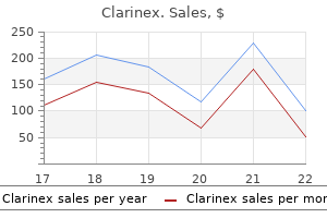
Cheap clarinex 5mg amex. Allergies Treatment & Cure Binaural Beats Meditation | All Kinds of Allergy Relief | Good Vibes.