

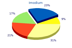
Lauren Grossman, MD, MS
- Clinical Instructor of Surgery
- University of Colorado Health Sciences Center
- Denver, Colorado
- Staff Physician
- Emergency Department
- Swedish Medical Center
- Englewood, Colorado
- Attending Staff Physician
- Department of Emergency Medicine
- Denver Health Medical Center
- Denver, Colorado
Placenta percreta: Balloon occlusion and embolization of the interior iliac arteries to scale back intraoperative blood losses gastritis definition wikipedia order 2 mg imodium otc. Temporary balloon occlusion of the frequent iliac artery: New approach to bleeding control throughout cesarean hysterectomy for placenta percreta gastritis symptoms belching buy imodium mastercard. Balloon-assisted occlusion of the interior iliac arteries in patients with placenta accreta/percreta gastritis diet dairy purchase imodium 2 mg on line. Placenta accreta: Comparison of instances managed with and with out pelvic artery balloon catheters gastritis disease definition purchase imodium overnight. Interventional radiology in ladies with suspected placenta accreta undergoing caesarean part gastritis gurgling buy generic imodium canada. Sciatic nerve ischaemia after iliac artery occlusion balloon catheter placement for placenta percreta gastritis journal articles cheap imodium amex. Prophylactic balloon occlusion of the interior iliac arteries to deal with abnormal placentation: A cautionary case. Pelvic umbrella pack for refractory obstetric hemorrhage secondary to posterior uterine rupture. Conservative management of placenta previa-accreta by prophylactic uterine arteries ligation and uterine tamponade. Systematic review of conservative management of postpartum hemorrhage: What to do when medical remedy fails. The triple-P procedure as a conservative surgical various to peripartum hysterectomy for placenta percreta. Placenta accreta encountered throughout dilation and evacuation in the second trimester. Diagnosis in the first trimester of placenta accreta with previous Cesarean part. Pelvic embolization for treatment of hemorrhage related to spontaneous and induced abortion. Scheduled hysterectomy for second-trimester abortion in a affected person with placenta accreta. Mifepristone and misoprostol for the administration of placenta accreta-A new various strategy. The main targets are to (1) decrease severe maternal morbidity associated to the placental illness, especially the quantity of blood loss (in turn, this decreases the chance of huge transfusion and coagulopathy in addition to operative injury, mainly bladder and ureteral damage, and its potential consequences such as vesicouterine fistula) and (2) try to preserve the choice of future pregnancies, figuring out that fertility is usually inextricably linked with societal status and shallowness. Four main types of conservative management have been described: (1) extirpative therapy,1 (2) expectant administration or leaving the placenta in situ,2 (3) one-step conservative surgery,three and (4) the Triple-P process. Leaving Placenta In Situ or Expectant Management Short- and Mid-Term Maternal Outcome this strategy consists of leaving the placenta in situ and ready for full resorption. It was first described mainly in France2 and initially was termed "conservative treatment of placenta accreta. This additionally will occur inside the placenta, and the placenta will progressively and spontaneously detach from the uterus and even adjoining organs by necrosis. These embrace intrauterine an infection, placental abscess, and even sepsis, as nicely as unpredictable large hemorrhage. Moreover, it requires long-term monitoring till complete resorption of the placenta happens. In apply, the precise place of the placenta is decided by a preoperative ultrasound. Postoperative antibiotic remedy (amoxicillin and clavulanic acid) is often administered prophylactically for 5 days to minimize the chance of infection, though efficacy is unsure. As with antibiotic treatment, none of these interventions has been confirmed to enhance outcomes. In France, the primary conservative treatment happened in 1993; the variety of procedures elevated steadily, particularly through the 2000s. Moreover, they have been from small case reports and case series from particular person tertiary-care establishments. Of the 26 women managed conservatively while leaving the placenta in situ without the use of extra therapies, 22 (85%) had a good outcome. Expectant administration failed in four (15%) of the sufferers who required hysterectomy due to severe hemorrhage or infection. Placenta accreta was identified in accordance with the following scientific and histologic criteria: (1) It was partially or totally inconceivable to manually remove the placenta with no discernable cleavage aircraft between all or a half of the placenta and uterus. Women treated with an extirpative approach or a planned cesarean-hysterectomy were excluded from this research. When placenta accreta was not suspected before supply, it was identified when it was impossible to detach the placenta by gentle manipulation, and conservative therapy was outlined as leaving half or all of it within the uterus. The examine included 167 circumstances of placenta accreta with 59% of placentas left partially in situ and 41% left completely in situ. Other rare morbidities included vesicovaginal fistula and arteriovenus fistula formation. Hysteroscopic resection and/ or curettage had been carried out to take away any remaining placenta in 25% with a median of 20 weeks (minimum: 2 weeks, most: 45 weeks). Notes: Data introduced as mean � standard deviation, or as median with interquartile range in parentheses, or as number of patients with percentages in parentheses. A main hysterectomy happened within the first 24 hours, whereas a delayed hysterectomy happened greater than 24 hours after supply. They found fifty seven cases of suspected placenta percreta that have been managed conservatively with the placenta left in situ. Hysterectomy was prevented in 60% of cases and 42% skilled major morbidity (including sepsis, coagulopathy, hemorrhage, pulmonary embolism, fistula, and arteriovenous malformation). Of the eight circumstances of placenta percreta with bladder involvement, conservative remedy was profitable in 6 circumstances (75%), and severe maternal morbidity occurred in 2 (25%). It also is critical that they comply with close follow-up monitoring in facilities with adequate tools and assets. The major downside of this strategy is that attempted removal of the placenta may cause severe bleeding with the danger of maternal hemorrhagic complications and hysterectomy. This can scale back the volume of placenta left within the uterus, potentially lowering the chance of bleeding and infection. These false-positive results may lead caregivers to carry out pointless radical or conservative surgical procedures that may also lead to possible complications. Thus, our current practice is to try and gently remove the placenta by cord traction solely in circumstances when the prognosis of placenta accreta is unsure. An instance could be a nulliparous girl with a history of curettage for whom ultrasound revealed intraplacental lacunae in a low-lying anterior placenta and no seen proof of placenta accreta through the cesarean. Some authors have proposed the use of methotrexate to hasten placental decision. In addition, methotrexate rarely causes severe harm such as neutropenia or medullary aplasia, even with a single dose in a young patient. Conservative Management of Placenta Accreta � Should preventive uterine devascularization be carried out in the absence of bleeding In reality, in a retrospective comparative research, the median delay for complete placental resorption was significantly shorter when women underwent an embolization (median = 17 weeks; q1011. Unfortunately, the rationale for embolization was not clearly reported by the authors. In our practice, we observe the patient in the hospital for eight days and administer prophylactic antibiotics for 5 days. Prior to discharge, the lady and her partner should be advised about the need for shut, long-term monitoring. The following signs require emergency medical consideration: hyperthermia, severe pelvic ache, foul-smelling vaginal discharge, and bleeding. The patient additionally should be suggested about the potential for abnormal and protracted vaginal discharge. There must be a multidisciplinary group available with the skills to handle complications 24 hours a day, 7 days a week. If the patient is asymptomatic, monthly visits are then carried out till complete resorption of the placenta. The visits embrace a medical examination (bleeding, temperature, pelvic pain), pelvic ultrasound (size of retained tissue), and laboratory display screen for an infection (hemoglobin and leukocytes, C-reactive protein, vaginal sample for bacteriological analysis). As mentioned above, in a French retrospective research, either hysteroscopic resection or curettage or both have been used to remove retained placenta in 29 (25. Nevertheless, no information concerning the reason for performing this process (due to pain, bleeding, and/or an infection, to hasten placental resorption, on maternal request, or systematically) was available. In a small cohort sequence of 23 girls with placenta left in situ for placenta accreta, 12 had undergone hysteroscopy under ultrasound guidance as a result of ache and/or bleeding with retained tissues. Complete removal (11/12) was achieved after one, two, and three hysteroscopic procedures in 5 (41. One delayed hysterectomy was performed after "failure" of the hysteroscopic procedure. The position of prophylactic hysteroscopy or the timing of it in asymptomatic ladies is unknown. A possible benefit of leaving the placenta in situ is to plan a delayed interval hysterectomy after partial involution of the placenta and decreased uterine vascularity. There had been eight ladies who had extreme intrauterine synechiae and have been amenorrheic. Of the 27 ladies who wanted more children, 3 girls had been making an attempt to become pregnant (mean duration: eleven. There is only one small retrospective examine comparing maternal end result following cesareanhysterectomy (n = 16) vs. As with delayed interval Conservative Management of Placenta Accreta 99 hysterectomy, the relative deserves of deliberate cesarean-hysterectomy and conservative administration will solely be elucidated via correctly designed clinical trials. Until such trials are accomplished, it appears reasonable to counsel ladies about planned cesareanhysterectomy and conservative management. A deliberate cesarean-hysterectomy could also be the most suitable choice if the patient has no need for more youngsters, is older, and/or multiparous. The primary steps of this uterine-sparing technique achieved through a median or Pfannenstiel incision are (1) vascular disconnection of newly formed vessels and the separation of invaded uterine from invaded vesical tissues, (2) efficiency of an upper-segmental hysterotomy, (3) resection of all invaded tissue and the whole placenta in a single piece, (4) use of surgical procedures for hemostasis, (5) myometrial reconstruction in two planes, and (6) bladder repair if essential. The indications for the 18 hysterectomies were segmental circumferential rupture greater than 50% (n = 13), coagulopathy (n = 2), an infection (n = 1), and uncontrolled hemodynamic instability (n = 2). The following issues were reported (mostly in group 2): lower ureteral injuries (n = 2), vesical fistula (n = 1), hematoma in the vaginal cuff (n = 1), and uterine an infection (n = 1). It is essential to note that the one-step procedure may be much less reproducible than conservative therapy because it requires a novel and specific surgical process. Successful use of the procedure by different teams and potential trials will ultimately clarify the merits of the one-step conservative remedy. It appears necessary to ensure that a 2-cm margin of myometrium is retained within the decrease lip of the uterine incision to facilitate closure of the myometrial defect. If the posterior wall of the bladder is concerned, placental tissue invading the bladder is left in situ to keep away from cystotomy. There was no statistical difference for the estimated mean blood loss (2,170 mL vs. One major complication (5%) occurred in a lady handled with Triple-P (right frequent iliac and exterior iliac artery thrombosis). The best-studied conservative approach is expectant care after leaving placenta in situ. Prospective trials are desperately wanted to assess the true dangers and benefits of conservative administration overall in addition to for each strategy. All authors contributed to the writing of the final version and to its revision for important mental content material, and all have seen and accredited the ultimate model. Prevention of postpartum hemorrhage and hysterectomy in sufferers with morbidly adherent placenta: A cohort study comparing outcomes before and after introduction of the Triple-P process. Clinical administration pointers for obstetricians and gynecologists: Postpartum hemorrhage. Active administration of the third stage of labour: Prevention and treatment of postpartum hemorrhage. Placenta Praevia, Placenta Praevia Accreta and Vasapraevia: Diagnosis and Management. Arteriovenous malformation identification after conservative management of placenta percreta with uterine artery embolization and adjunctive therapy. Arteriovenous malformation following conservative remedy of placenta percreta with uterine artery embolization however no adjunctive therapy. Life-threatening neutropenia following methotrexate therapy of ectopic being pregnant: A report of two instances. Fertility and pregnancy outcomes following uterine devascularization for extreme postpartum haemorrhage. Predictors of failed pelvic arterial embolization for severe postpartum hemorrhage. Planned caesarean within the interventional radiology cath lab to allow quick uterine artery embolization for the conservative therapy of placenta accreta. Uterine necrosis following pelvic arterial embolization for post-partum hemorrhage: Review of the literature. Conservative administration of morbidly adherant placenta- A case report and review of literature. Conservative management of placenta accreta: Hysteroscopic resection of retained tissues. Medical and surgical therapy of placenta percreta to optimize bladder preservation. Planned caesarean hysterectomy versus "conserving" caesarean part in sufferers with placenta accreta.
Generally gastritis diet vegan generic imodium 2 mg otc, gentle amounts of dystonia gastritis diet for children buy 2mg imodium with mastercard, bradykinesia gastritis symptoms diet buy imodium 2mg overnight delivery, and rigidity of limbs coexist with comparatively extra prominent chorea gastritis diet ����� buy 2 mg imodium fast delivery, which itself reaches a peak in mid-stage disease gastritis symptoms ayurveda order 2 mg imodium, after which this balance reverses as illness progresses gastritis neurological symptoms discount 2 mg imodium with visa. In average and superior phases, lack of both involuntary and voluntary motion occurs with irregular fastened postures, particularly of the trunk or neck, that are usually dystonic in origin. Flexion contractures can ensue reflecting overriding spasticity and rigidity and representing more florid degeneration of corticospinal tracts. The exception is the Westphal, akinetic-rigid variant, the place rigidity and bradykinesia prevail and tremor and chorea are not often seen. Axial rigidity appears to be extra significantly related to disability and lack of ambulation than chorea [20]. Motor impersistence can be elicited by asking the patient to keep a completely protruded tongue for 10 seconds. Patients show important postural management deficits when replicating motor abilities employed in actions of day by day living, emphasizing the functional impairment these symptoms confer [22]. At superior levels of disease, fixed dystonic posturing of head, trunk, or limbs can happen, necessitating acceptable secondary preventative measures. Eye actions the broken pursuit movements and slowed saccades (vertical worse than horizontal) seen early on the disease worsen, requiring head actions or eye blinking to provoke, and at the advanced phases an entire supranuclear gaze palsy is feasible [23]. A characteristic signal is gaze impersistence with issue fixating on an object. These oculomotor abnormalities are prone to represent disruption to the fronto-striatal control of eye actions [24]. Patients report difficulty in swallowing solid and liquids and can also choke sometimes, emphasizing the importance of speech and language therapists, dieticians, and considered investigation in the form of videofluoroscopy. In the later levels, progressive swallowing and talking impairment gives way to anarthria and mutism. Continence Urge incontinence with related frequency and urgency is probably essentially the most generally reported criticism, normally occurring in mid-stage disease. A research of six patients revealed four with detrusor instability while two had a normal study suggesting a non-organic disturbance. A characteristic urodynamic pattern consisting of choreiform movements of the pelvic flooring musculature throughout filling with selective suppression of choreiform contractions within the perineum throughout detrusor contraction was noticed although this has never been adopted up [26]. In a newer research, cystometry performed in 12 manifest patients and 1 premanifest affected person demonstrated detrusor�sphincter dyssynergia, detrusor overactivity, and decreased detrusor capability [27]. Hypothalamic-pituitary dysfunction does undoubtedly play a job leading to sleep fragmentation and sleep-wake cycle reversal. Depression and apathy may contribute additionally by way of a loss of daytime stimulation and exercise. Involuntary actions can hinder getting off to sleep in addition to contribute to middle-of-thenight insomnia. Occasionally this may manifest or certainly be supplemented with a outstanding behavioral change corresponding to aggression or alternatively withdrawal and alter of character. Symptoms may be present from the premanifest stage, with one research showing that 38% of patients in the premanifest stage had gentle cognitive impairment [34]. Deficits in processing velocity, visuospatial processing, time estimation and timekeeping, learning, and working memory are also seen in this group. Correspondingly, they report difficulty in selecting up new tasks but retrieval of taskspecific recollections can be affected. There can also be dependable proof of olfactory impairment in both premanifest and manifest sufferers [35, 36]. It is obvious also that cognitive and behavioral signs place the best burden on carers and correlate with functional decline and institutionalization [37]. However comprehension is believed to be properly preserved even in late stages of the illness. Communication, however, can be impacted upon by diminished processing speed and loss of coordinated oro-motor function with respiration [36]. Current apply might embrace the Verbal Fluency, Symbol Digit, Stroop, and Category fluency as nicely as Trails A and B and the Hopkins Verbal Learning Test, as derived from a quantity of years of observational knowledge [36, 38, 39]. Symptom Irritability [41] Apathy [41] Anxiety [41] Depressed mood [41] Obsessive and compulsive [41] Bipolar symptoms [42] Psychotic [41] Prevalence (%) 38�73% 34�76% 34�61% 33�69% 10�52% 5�10% 3�11% 252 Neurodegeneration tability, apathy, obsessive-compulsive signs, psychosis, and substance abuse amongst others. Similarly, in premanifest sufferers, psychiatric options are famous on scales of depression, obsessivecompulsiveness, anxiousness, and psychosis, although often not enough to meet established diagnostic standards. Over a 2-year follow-up of mutation carriers, 15% of sufferers without a pre-existing psychiatric illness at baseline developed a dysfunction, most frequently melancholy, which was seen in 64% of topics [40, 42]. It is obvious in scientific follow that particular person, so-called "subsyndromic" features of several psychiatric illnesses may be present in the identical subject. Patients with obsessive and compulsive symptoms show a better prevalence of melancholy, suicidal ideation, and psychotic phenomena, whereas others have isolated low mood or obsessive, repetitive behaviors without the accompanying compulsions, rendering their medical administration difficult [43]. Further on, transitioning from premanifest to manifest disease, adopted most poignantly by progressive lack of function, standing, and independence, has the potential to aggravate or indeed unmask psychopathology. In a survey of 2835 sufferers, 40% had signs in keeping with despair, whereas 50% had beforehand acquired remedy for melancholy [45]. Although statistics on suicide charges have been somewhat conflicting, patients have a lifetime threat 4�6 instances higher than the overall inhabitants. A meta-analysis of mortality associated with mental well being problems which included 4171 gene carriers revealed that 17. The threat appeared best in those that had been perimanifest, experiencing the primary, significant losses in perform, and struggling with depression and impulsivity [2]. Irritability Alongside depressive signs, irritability is often encountered either as an isolated presentation or as part of melancholy and infrequently psychosis. A working definition of irritability is lack of temper at a much decreased threshold compared to the overall inhabitants, at seemingly innocuous stimuli. This could be difficult by aggressive outbursts, which may vary from verbal aggression with cursing, to verbal threats of physical violence, and eventually to bodily damage itself. Irritability results from a large number of factors, together with frustration at declining operate and cognitive overload within the setting of diminished psychomotor ability and planning. Irritability appears to be as frequent as despair, if not more so, and occurs all through. Differentiation of sure aspects of the presentation corresponding to disinhibition, irritability, or hypersexuality, from frontal symptomatology may be tough. Moreover, the basic triad of altered fluctuating temper, a grandiose thought content material, and overactivity is incessantly lacking in these patients. In more extreme types, delusions and hallucinations can happen, whereas at the different end of the spectrum, rapid biking of mood without other overt bipolar indicators could be seen. The traditional psychotic presentation includes paranoid and stuck concepts with accompanying aggression, irritability, and poor impulse control [48]. Delusional and schizophrenia-like psychotic states are rare but can characteristic delusions centered on erotomania, bodily decay, parasitosis, and third-party auditory hallucinations. Psychosis can also present within the later phases of the illness, the place it can be masked by the concomitant cognitive decline. However, repetitive and perseverative behaviors and speech content are sometimes seen as part of the frontal dysexecutive syndrome. In support, following a cross-sectional study, lack of power and apathy was discovered to affect greater than 75% of sufferers. Correlation was famous with the length of illness and though there was coexistence of irritability and melancholy, a subsequent research demonstrated that apathy was independent of depressive pathology [44, 50]. When depressed patients were excluded, male intercourse, diminished world efficiency, and greater neuroleptic and benzodiazepine burden were independently correlated with apathy [51]. Weight loss Unintended weight reduction regardless of elevated appetite and calorie intake, a minimum of in early illness, culminates in the cachexia of superior levels. In certain sufferers startling weight reduction can be seen due to a mixture of dysphagia and bodily difficulties in using cutlery and going out to buy meals. Weight loss can be exacerbated by neuropsychiatric aspects corresponding to despair and lack of motivation and desire to eat, in addition to the recognized catabolic nature of the illness. This may be compounded by lack of hypothalamic somatostatin and orexin neurons [57]. Xerostomia, along with loss of lingual dexterity and swallowing difficulties, may also contribute to mechanistic impairment of the feeding course of [58]. Importantly, it has been famous that patients with a higher physique mass index have a slower price of development, and accordingly medical follow recommends weight upkeep with high-calorie diets [60]. In a big retrospective study, heart illness was the second leading cause of death [69]. Multiple contributory factors including the disease itself, its complications, and the medications employed in its management are implicated. In this setting, occult subdural hematomas from unwitnessed falls and head injuries ought to always be considered, as should cerebrovascular disease. Poor nutritional consumption can lead to quantity depletion and metabolic disturbances; urogenital dysfunction might predispose to urinary tract infections, and oromotor dysfunction to pneumonia. It ought to be famous that, as with different neurological ailments, in the presence of generalized sepsis even beforehand intermittent chorea can appear florid and any subclinical features similar to cognitive impairment, neuropsychiatric disease, and steadiness difficulties may be magnified. The cumulative effects of diminished saliva manufacturing, poor meals clearance, and decreased capacity to brush teeth mean that oral hygiene could also be compromised and therefore dental health must be rigorously maintained as illness advances. In superior phases the following are important: prevention of strain sores, flexion contractures, and self-injury; good pores and skin and nail care; padded wheelchair provision; and prevention of deep vein thrombosis. Finally, although a wide range of signs exist, clinicians should guard towards mechanically attributing any new symptom to disease progression, as other frequent medical situations can and do happen in this inhabitants. Premanifest phase of disease Classically, analysis has relied heavily on the presence of motor symptoms. It is only relatively lately that large multicenter observational studies have formally confirmed what clinicians have long suspected � the existence of a premanifest section, characterized variably by evolving subtle motor deficits, and at instances vital cognitive or psychiatric signs and indicators, occurring nicely before overt onset of motor symptoms and indicators. This period has been variously termed pre-symptomatic, pre-disease, or prodromal; a universally agreed taxonomy is missing, although right here we use the term premanifest. In terms of presenting symptoms, refined motor features similar to slight chorea can be present. Early instability of stability and posture can be considered a premanifest motor signal as balance and posture have been objectively demonstrated to be impaired in premanifest subjects versus controls [75]. Abnormalities in easy pursuit and saccadic eye movements are arguably some of the dependable early motor indicators. Alongside the abnormalities on the Luria take a look at and presence of chorea, they seem to correlate most carefully with an escalating probability of scientific analysis. Elevated total motor scores at baseline had been related to higher genetic chance of illness prognosis within the close to future and smaller striatal volumes [76, 77]. In a longitudinal sub-study, 29 premanifest topics and 43 non-carrier controls underwent clinical testing and an intensive neuropsychological take a look at battery addressing international cognitive function, reminiscence, language, and executive perform. In symptomatic phrases, sufferers might display change in conduct similar to aggression, apathy, and lack of interest or report poor performance at work, notably during multitasking. Emotional recognition, which requires interpretation of facial expressions and verbal intonations in addition to body language consultant of worry, sadness, or disgust, was additionally proven to be impaired in premanifest topics [79]. Although there were initially confounding leads to the literature, methodological enhancements have shown elevated frequency of psychiatric symptoms in premanifest subjects suggestive of despair, anxiousness, and obsessive-compulsive dysfunction as compared with gene-negative people. Indeed, those with higher motor scores appeared to have higher ranges of psychiatric morbidity, even though the vast majority of subjects had been estimated to be greater than 10 years from predicted prognosis [80]. In the premanifest group, regardless of significant declines in regional and total brain volumes, few useful variables confirmed important 24-month change in contrast with controls; whole motor rating, emotion recognition, and speeded tapping had been exceptions. Premanifest individuals with development exhibited higher rates of brain atrophy and deterioration on some quantitative motor tasks compared with other premanifest individuals [38, 39, 82]. The early stage represents the point at which sufferers have minimal limitation, are often in a position to keep employment without compromise, and proceed to stay independently. Minor involuntary actions, psychiatric illness, and some problem when multitasking may be reported. The major difference at the center stage is the lack of employment whereas still maintaining actions of daily residing. Chorea is normally at its peak at this stage, with lack of fine motor management, increasing falls, and abnormal mobility and gait. At the late disease stage, complete immobility is present as individuals are bedbound and require help in all activities of every day dwelling. Profound dysarthria additionally happens to the point of severe communication difficulties. Chorea is almost universal however the course is milder and slower with gentle cognitive and psychiatric illness. Late-onset illness is usually related to repeat sizes of forty, although lengths of as much as forty eight have been recorded. Often a household history can be lacking, which can replicate enlargement of an intermediate vary allele [85, 86]. In the most important case collection of juvenile sufferers, over half skilled symptom onset beneath the age of 14 while 1 in 10 instances occurred before the age of 1 year. Under the age of 10, developmental delay specifically in speech and language may be distinguished and will manifest as failure to progress at college. Behavioral adjustments, learning difficulties, rapid cognitive decline, psychiatric disease, and parkinsonian motor options with predominating rigidity and bradykinesia have been seen in 50% of instances, as well as dystonia and ataxia. The vary of psychiatric disease can embody drug and alcohol abuse as properly as consuming problems [88]. The most typical signs reported by the households have been speech difficulties, dysphagia, stiffness/spasticity, sleeping problem, pain, and behavioral issues [89]. In distinction to the grownup form, chorea itself is uncommon and early oropharyngeal dysfunction seems extra generally in juvenile cases.
C gastritis symptoms back pain cheap imodium 2mg amex, Following distinction administration moderate gastritis diet buy imodium 2mg line, the lesion is shown to have stable (arrowhead) and cystic (arrows) parts gastritis symptoms lower abdominal pain cheap imodium 2mg fast delivery, a not unusual feature of schwannomas gastritis burning stomach cheap imodium 2mg with amex. Lateral meningoceles may be related to wormian bones gastritis diet ������� purchase cheap imodium line, malar hypoplasia chronic gastritis reversible cheap 2mg imodium mastercard, developmental delay, quick stature, and spontaneous intracranial hypotension. These diseases are characterized by concentric proliferation of Schwann cells, interspersed with collagen, in response to a number of episodes of demyelination and remyelination. Patients current with a wide selection of signs and signs relying on the particular illness. Guillain-Barr� syndrome is an autoimmune disorder that can happen following viral or bacterial an infection (most commonly Campylobacter jejuni infections). Although Guillain-Barr� syndrome favors motor neurons, sensory and autonomic nerves can additionally be affected. Patients typically present with symptoms ranging from ascending weakness to flaccid paralysis. Several subtypes exist, together with acute inflammatory demyelinating polyradiculopathy, axonal subtypes, and regional syndromes (including the Miller-Fisher variant by which ataxia and areflexia are the dominant features). Additionally, paraneoplastic syndromes have been reported to enlarge cranial and peripheral nerves produced by a microvasculitis related to inflammatory changes. There is a female predominance and a excessive incidence (as excessive as 60%) of malignant transformation, especially in male patients. Lesions present as a big expansile mass, with fatty, cystic, and solid components, and may even be hemorrhagic. Extradural Lesions the vertebral bodies are essentially the most frequent web site of metastatic illness to the backbone. A, Markedly thickened lumbar nerve roots are current on this affected person with Charcot-Marie-Tooth disease. B, In this identical patient, even the ventral and dorsal nerve roots arising from the cervical twine are massively enlarged (arrows) as are the exiting nerve roots inside the neural foramina (arrowheads). Chapter 16 Nondegenerative Diseases of the Spine 579 sign intensity compared with the normal fatty marrow with lytic lesions, or may seem as lower sign depth with sclerotic lesions. Common cancers associated with spinal metastasis embody breast, prostate, lung, and kidney. Other lesions include Ewing sarcoma, neuroblastoma, melanoma, lymphoma, leukemia, a quantity of myeloma, and sarcoma. Pelvic lesions metastasize through the epidural plexus of veins (Batson plexus) to the thoracolumbar our bodies. Multiplicity of lesions within the vertebral column should raise suspicion for metastatic illness. Oftentimes, the primary tumor is known and metastatic illness is suspected based mostly on affected person symptomatology. In these instances, you will need to not solely determine metastatic lesions to bone but to also assess for problems of metastatic disease. This includes an evaluation for pathologic fracture, extraosseous soft-tissue extension into the perivertebral and intraspinal spaces, cord compression, and cord sign changes. Osteoporotic Compression Fractures Osteoporosis is characterised by decreased bone mass. It is associated with compression fractures that are spontaneous or related to minimal trauma. Osteoporosis is the commonest explanation for compression fractures and is particularly observed in aged women. The ache from an acute fracture often lasts 4 to 6 weeks and is associated with beautiful ache at the fracture web site. A, Far lateral image from sagittal quick tau inversion recovery sequence reveals marked enlargement of the cervical nerve roots as they exit the neural foramina (arrows). The presence of all bone fragments from the original cortex (puzzle sign) is each very constant and fairly specific for benign disease. It is attention-grabbing that sclerosis, marked comminution of cancellous bone of the vertebral body, Schmorl nodes, or fracture of a pedicle have low specificity for separating benign from malignant illness. Both may involve a number of vertebral bodies and be related to soft-tissue lots. A paraspinal mass is more prone to be neoplastic; however, soft-tissue hematoma associated with benign compression fracture could make the excellence difficult. In the presence of other metastatic lesions within the backbone, the collapsed vertebra more than likely outcomes from metastatic illness. Fatty filum in a special affected person seen as T1 hyperintensity (E) and T2 hyperintensity (F), paralleling the course of the cauda equina (arrows). In the case of a single collapsed vertebral body in a patient with or without metastatic illness, a repeat examination in four to 6 weeks is beneficial. Naturally, the patient will have a full workup for occult malignancy, including bone scan to determine whether or not there are different lesions. The list of illnesses that may produce vertebral compression includes metastases, osteomyelitis, congenitally poor vertebral endplates, benign and malignant primary vertebral neoplasms, osteomalacia, hyperparathyroidism, hemochromatosis, myeloproliferative disorders, and trauma. Neural Crest Tumors Tumors of neural crest origin happen in infancy and come up from the sympathetic plexus. C, Lateral backbone radiograph in a different affected person with metastatic breast most cancers reveals in depth osteoblastic metastases. These paravertebral lots, which often occur within the thoracic region, can lengthen via the neural foramina to compress the thecal sac (yet extra dumbbells. When evaluating paraspinal masses, extramedullary hematopoiesis should probably cross your mind. This course of happens when normal erythropoiesis within the marrow fails because of problems in hemoglobin manufacturing (like sickle cell disease) and myeloproliferation (like myelofibrosis or chronic myelogenous leukemia), prompting purple cell manufacturing exterior the marrow compartment. Hemangioma Hemangiomas are widespread benign lesions of the vertebral body and are most often incidental findings. More than 50% of solitary hemangiomas occur in the backbone and so they have been found in approximately 12% of spines at post-mortem. There is a 2:1 female/male predominance, with most lesions found in the decrease thoracic and lumbar vertebrae. These lesions most often current within the vertebral bodies as round geographic lesions but hardly ever can be extensive, replacing the whole vertebral body with extension into the pedicles, arches, and spinous processes. The vertical striations symbolize vascular channels interspersed with thickened trabeculae. While benign, vertebral bodies with hemangiomas can have compression fractures or epidural extension and compromise the spinal canal or neural foramen. Problems occur when these lesions compress the spinal cord (myelopathy) or when they become hypervascular and produce venous hypertension. Most hemangiomas are asymptomatic; nevertheless, ache is associated with progress (especially in ladies during pregnancy) and compression fracture. Very not often, they are often aggressive and show extraosseous extension, producing myelopathy from wire compression. Otherwise patients could have to go to biopsy ("bloody hell") or undergo follow-up imaging to assess for interval change. Chordoma Chordomas are slow-growing, locally invasive neoplasms derived from remnants of the notochord, with approximately 50% originating within the sacral area and 15% affecting the spine (particularly the cervical region). In the spine, the tumor mass is related to lytic lesions of the bone, at instances with a sclerotic rim and calcification. Eosinophilic Granuloma Eosinophilic granuloma (part of the Langerhans cell histiocytosis spectrum) affects children and when seen within the spine predominantly entails the vertebral body. There could also be associated soft-tissue extension from the vertebral physique posteriorly into the vertebral canal or anteriorly into the prevertebral tissues. Osteochondroma these lesions often arise from the posterior elements and commonly involve the thoracic or lumbar vertebrae. They are commonly found in youngsters where they grow and infrequently trigger neurologic signs. Paget Disease Paget disease generally includes the backbone and produces enlargement of all of the vertebral elements. An early lytic part has also been described (with osteoporosis circumscripta when seen within the skull). Involvement is associated with back and neck ache and neurologic dysfunction associated with side arthropathy, lateral recess syndrome, and stenosis of the spinal canal. It is difficult at instances to distinguish from osteoblastic metastatic illness because Paget illness is polyostotic in greater than two thirds of circumstances. There is the potential of malignant transformation to osteogenic sarcoma in roughly 10% of cases: the rate of malignancy is 20 instances higher in pagetoid bone than in nonpagetoid bones. The look of osteogenic sarcoma is variable, depending on whether or not the lesion is lytic, blastic, or blended. Both symptomatic and asymptomatic sufferers might reveal these patterns early in the midst of the illness. These lesions have some characteristics that may help distinguish them from one another. Giant cell tumors are lytic lesions found commonly in the sacrum and vertebral our bodies; tumor margins may be skinny or dehiscent and soft-tissue component of the mass may be present. C, Contrast that with this case, where there are multiple bilateral paraspinal plenty. Before you jump into that neural crest tumor differential, you should know that is an grownup affected person with thalassemia. Vertebral compression fractures occur in 50% to 70% of sufferers and spinal twine compression in 10% to 15% of those circumstances. Rarely, a quantity of myeloma can contain the leptomeninges with enhancement of the nerve roots. Consider myeloma in a patient with diffuse osteopenia and compression fractures (particularly male patients). Remember that myeloma is amongst the illnesses that might be tough to detect with bone scans. A solitary plasmacytoma may be indistinguishable from a lytic metastasis and is a harbinger of a quantity of myeloma. Vertebroplasty this system has been used to treat pain ensuing from both benign and malignant vertebral physique collapse. Complications end result from leakage of cement into perivertebral veins resulting in compression of the spinal twine or nerves or pulmonary embolism. There is a circumscribed lytic region in the right lamina on this computed tomography scan with bone windows. In the central portion of the hypodense region (arrow) is a punctate area of excessive density. Kyphoplasty is a associated technique to vertebroplasty where a balloon is blown up in the vertebral physique after the insertion of the needle via the pedicle. Sagittal reconstructions from computed tomography lumbar backbone reveals loss of peak at L3 (arrows) with lytic look within the medullary compartment together with posterior parts (arrowhead). This lack of height, vertebra plana, is attribute of eosinophilic granuloma involving the backbone. Epidural Lipomatosis Epidural lipomatosis can happen from a selection of totally different causes together with obesity, steroid use (usually after prolonged use of oral steroids), and Cushing syndrome. The fatty tissue is seen most frequently within the posterior epidural area and may contribute to spinal stenosis and may produce significant wire or cauda equina compression. Treatment includes weight loss and cessation of steroid use, relying on the cause. The medical presentation contains profound impairment of bowel and bladder operate, loss of perineal sensation, and moderate impairment of sensory and motor operate of the decrease extremities. Ischemia to the conus could end result from poor collateral provide after occlusion of the dominant blood supply (artery of Adamkiewicz). Infarction may be the outcome of problems related to the descending aorta, corresponding to atheroma, aortic surgical procedure, and dissecting aneurysm. Other causes embrace vertebral occlusion or dissection, arteritis, vascular malformations, being pregnant, hypotension, sickle cell anemia, tuberculosis, meningitis, arachnoiditis, vascular malformation, diabetes, degenerative illness of the spine, and disk herniation with spinal artery damage. Careful consideration ought to be paid to the aorta for aortic dissection or aneurysms as a cause. If a technically enough diffusion-weighted sequence could be carried out within the spine, will probably be brightly constructive like most infarcts and seal the deal. Treatment of spinal vascular malformations is dependent on many factors, including age, malformation kind, and neurologic situation, and consists of embolization, surgery, or a combination of the two. Blood provide to the malformation is very important in figuring out whether to proceed with embolization or to perform surgery. The differential diagnosis of subarachnoid hemorrhage and hematomyelia is offered in Box 16-11. While the imaging findings carry a differential diagnosis, the clinical context of abrupt loss of sensation and weak point abruptly following aortic aneurysm restore ought to make the analysis of wire infarct a no brainer. C, Diffusion-weighted imaging from another patient reveals excessive signal at the stage of wire infarct (arrow) relative to the traditional wire. This malformation, whose draining veins are most incessantly found on the dorsal side of the lower thoracic wire or conus medullaris, generally has the eponym Foix-Alajouanine syndrome attached to it, to describe the myelopathy secondary to venous hypertension within the twine. In approximately 85% of circumstances, a single radicular artery with systemic strain is identified draining instantly into spinal pial vein(s). However, there are cases with many arterial feeders originating from either single or a number of ranges that could be both unilateral or bilateral. The systemic stress in the spinal veins initially dilates these vessels with subsequent kinking and poor venous drainage. This leads to venous hypertension defined histopathologically by stasis, edema, ischemia, and resulting in swelling and subsequent infarction of the spinal twine. Complaints begin with an insidious onset of lower extremity weak spot or sensory modifications, associated with nonradiating ache starting in the caudal spinal segments and progressing superiorly. There is a propensity for these to occur in males in their fifth or sixth decade of life.

Mesenchymal Meningeal Tumors As famous in Box 2-2 there are a variety of mesenchymal tumors that may affect the meninges gastritis diet ������� purchase 2 mg imodium. These are all relatively rare lesions gastritis diet and treatment buy generic imodium from india, which may have either osseous (osteoma prepyloric gastritis definition generic 2 mg imodium amex, osteosarcoma gastritis diet �������� purchase 2 mg imodium with mastercard, etc gastritis diet �������� order imodium overnight delivery. Hemangiopericytoma these tumors hcg diet gastritis 2 mg imodium, derived from smooth muscle pericyte cells of Zimmerman across the capillaries of the meninges, are extra aggressive than most meningiomas, have a higher price of recurrence, and might metastasize. They are likely to be giant (over four cm in size), lobular, extraaxial supratentorial plenty. Melanocytic Lesions Within this category one finds diffuse melanocytosis, melanocytoma, neurocutaneous melanosis, and malignant melanoma. These diagnoses are tough to distinguish from metastatic melanoma to the dura. The melanocytoma is the most common of the lot and is seen in adults, whereas melanocytosis is more of a pediatric dysfunction. Although the latter is a diffuse course of, the former normally presents as a posterior fossa mass. Malignant melanoma of the meninges could bleed or unfold from the dura to the adjacent nerves, brain, or cranium. Prognosis is poor as is malignant melanoma of the meninges with distant metastases. Tumors of Neurogenic Origin Schwannomas the intracranial neurogenic tumors (schwannomas, neurofibromas), are similar in appearance. Note that, relying on the degree of Antoni A and B tissue within schwannomas, the signal depth of these lesions may be brighter than or could directly simulate that of meningiomas. Other terms used for schwannomas embody neurilemmomas and neurinomas, but the most accurate time period is schwannoma. Chapter 2 Neoplasms of the Brain 45 the excellence between meningiomas and schwannomas is a common one which radiologists should make on the cranium base. Nonetheless, regardless of involving the vestibular, not cochlear, nerves, the patients present with listening to loss. Peritumoral edema could also be seen in a single third of instances, normally within the larger schwannomas. Hiryu has a cyst (C) related together with his solidly enhancing (T) tumor nodule on his enhanced T1-weighted image. A, Left intracanalicular vestibular schwannoma: Post gadolinium enhanced T1-weighted coronal scans on affected person Ena Ken Al present an intracanalicular mass (arrow) on the left facet. Patient Sister Na Duhl has a brightly enhancing mass with both a cisternal (black arrow) and intracanalicular (arrowheads) portion. For these circumstances with progressive, nodular, or masslike enhancement, suggest recurrence. Jugular schwannomas more commonly develop intracranially than extracranially and sometimes easily erode the jugular foramen. The border of the bone is scalloped versus the paraganglioma that has a way more irregular and nonsclerotic margin. Schwannomas compress the jugular vein, whereas paragangliomas (glomus jugulare tumors) invade the vein. Whether the schwannoma arises from one cranial nerve or the subsequent, the imaging look is similar. Neurofibromas Neurofibromas, strictly speaking, refer to the tumors related to neurofibromatosis. The pores and skin and subcutaneous tissues are affected more typically than peripheral nerves; spinal nerve neurofibromas are rare and cranial nerve involvement is rare. These lesions contain Schwann cells, perineural cells, and fibroblastic cells and should happen in a plexiform aggressive subtype, which appears as a network of diffusely infiltrating masses. A, Sagittal T1-weighted image in patient Case Ewell Mysse reveals a markedly thickened descending portion of the facial nerve (arrows). B, the course of this enhancing mass suggests a facial nerve schwannoma by virtue of its tympanic section and descending portion on this coronal post-contrast T1-weighted picture (asterisk). Hedd that programs alongside the pons to the Meckel cave and the best cavernous sinus (normal Meckel cave on left with arrow). With a tumor like this check the ipsilateral pterygopalatine fossa for spread (V-2) and muscles of mastication for atrophy (V-3). Plexiform neurofibromas, due to their extensiveness, may be distinguished from neuromas and schwannomas. They are generally found within the extremities or within the soft tissues of the top and neck. Neuromas By strict pathologic definition, neuromas refer to a posttraumatic proliferation of nerve cells quite than to a real neoplasm. They are often seen in the cervical spine when nerves are avulsed, or in an operative bed. Unfortunately, in the widespread vernacular most people imply "schwannoma" after they say "neuroma". Breast carcinoma is the most common neoplasm to be associated with purely dural metastases. In kids, dural metastases are mostly related to neuroblastomas and leukemia. These tumors are additionally well-known for lodging within the cranial sutures, widening them in an toddler. Inflammatory lesions which will simulate dural metastases include granulomatous infections (mycobacterial, syphilitic, and fungal), Erdheim Chester disease, sarcoidosis, and Langerhans cell histiocytosis. When the method is diffuse we use the time period "sugar-coating" of the subarachnoid house because the whole pial surface is studded with granules. Lymphoma will nearly always improve (unless the neurosurgeons are reluctantly treating it with large doses of steroids). Include meningioma, sarcoidosis, plasmacytoma, and other dural metastases in the differential prognosis. A, the gadolinium-enhanced scan seems identical to one would expect for a meningioma with marked enhancement and a dural-based lesion. The irregular margins (saw-toothed appearance) suggesting pial unfold can be funky for a meningioma though. B, Even the T2-weighted picture shows low sign that simulates a meningioma or lymphoma or sarcoidosis. Postgadolinium coronal scan in affected person Hope Anna Prere reveals a excessive left frontal intraparenchymal metastasis, which demonstrates dural invasion (arrows) alongside its superomedial margin. Edley show a lesion centered on the calvarium with extracalvarial, dural, and intraparenchymal invasion (open arrows). Clinically, the patients present with a number of cranial neuropathies, radiculopathies, and/or mental standing changes secondary to hydrocephalus or meningeal irritation. Postcontrast T1-weighted image reveals contrast-enhancing nodules (arrows) in the roof of the lateral ventricle and within the superior vermian cistern. In children, 80% happen on the trigone and/or atria of the lateral ventricles; in adults they may also be seen within the fourth ventricle. If you also embrace adults, 43% are located within the lateral ventricle, 39% in the fourth ventricle, 11% within the third ventricle, and 7% in the cerebellopontine angle cistern. Lately, the obstructionists have gained favor over the overproductionists so far as the reason for hydrocephalus. Calcification happens in 20% to 25% of choroid plexus papillomas, and hemorrhage in the tumor is seen even more frequently than calcification. Between the calcification, circulate voids, and/or hemorrhage, the tumor might have a very heterogeneous appearance, generally with a salt-and-pepper appearance from vessel supply. Lexus-Goombah has a lot of peripheral high signal on fluid-attenuated inversion recovery, signifying edema of the parenchyma. The voxel placed for magnetic resonance spectroscopy yielded high ranges of choline-containing compounds and a whole absence of creatine and the neuronal/axonal marker N-acetyl aspartate. Choroid Plexus Hemangiomas Choroid plexus hemangiomas are benign neoplasms of the choroid plexus normally seen within the lateral ventricle. In this syndrome choroidal hemangiomas ipsilateral to the leptomeningeal vascular malformation could additionally be present (see Chapter 8). Choroid Plexus Xanthogranuloma Another benign situation of the choroid plexus is the xanthogranuloma. This incidental lesion might have fats density/ depth inside it and is also centered on the glomus of the trigone. Other Intraventricular Masses (Excluding Meningiomas) Ependymal Tumors Ependymomas are considered one of a variety of ependymal tumors (Table 2-4) which will occur all through the brain and spinal twine. Although ependymomas usually present earlier than age 10, a second ependymoma peak in the fourth and fifth decade of life is seen. Posterior fossa ependymomas are usually associated with the fourth ventricle, though lesions arising primarily in the foramina of the fourth ventricle do happen and may present as masses outside the fourth ventricle (Table 2-5). Although it may appear a paradox, 20% of ependymomas come up intraparenchymally, often in the supratentorial house (thought to be due to ependymal rests left over from radial glial migration). There is a 50% 5-year progression-free survival rate with posterior fossa ependymomas. Ependymomas have a higher incidence of calcification (40% to 50%) than different posterior fossa pediatric neoplasms. The lesions reveal mild enhancement, to a lesser diploma than the medulloblastomas. Hydrocephalus is normally current because of blockage of the fourth ventricular outflow. They often come up as midline lesions that fill the fourth ventricle without displacing it. A classic appearance of a posterior fossa ependymoma is a calcified fourth ventricular mass that extends through and widens the foramina of Luschka and Magendie. When seen within the cerebral hemispheres, the lesions are bigger and are cystic 50% of the time. Ependymomas are another instance of mind tumors which have a high incidence of subarachnoid seeding, and the use of contrast material is essential to the detection of subarachnoid spread. Subependymoma Subependymomas are variants of ependymomas that contain subependymal neuroglia. They come up intraventricularly or periventricularly and are frequently multiple at post-mortem. Lateral ventricular subependymomas often come up in patients older than 10 to 15 years. A, this mass in affected person Dru Batluk fills and expands the fourth ventricle and is heterogeneous on T2-weighted image. B, Note how the lesion oozes by way of the foramen of Magendie (arrowhead) to squirt into the cervicomedullary junction. They are hardly ever seen in the spinal canal as intramedullary or extramedullary intradural masses. Intraventricular neurocytomas hemorrhage more incessantly than oligodendrogliomas, which can suggest that prognosis. Immunohistochemical markers for synaptophysin can distinguish intraventricular oligodendrogliomas from neurocytomas. This is finest for a subependymoma as a end result of a neurocytoma, meningioma, and choroid plexus papilloma will improve. Nonneoplastic "Tumors" Epidermoids There is some confusion concerned with placing epidermoids and dermoids right into a "tumor" chapter because these are congenital epidermal inclusion cysts and dermal inclusion cysts. Teratomas, usually lumped in the same class, however, are true neoplasms of multipotential germ cells. Epidermoids are additionally called congenital cholesteatomas and, like acquired ones within the temporal bone, have a pearlywhite appearance. Men and women are affected equally, with a peak incidence in the 20- to 40-year age vary. Extradural epidermoids are nine instances much less common than intradural ones and arise intraosseously within the petrous bone and the temporal bone. Sometimes epidermoids are hard to distinguish from arachnoid cysts, notably within the cerebellopontine angle cistern (Table 2-7). A, Axial enhanced fluid-attenuated inversion restoration scan reveals the left lateral ventricular mass in patient Rush Mityo Oh Arr hooked up to the septum pellucidum (arrow). Note the scalloped margin to the lesion at the pons-medulla-cervicomedullary area. E, Calvarial epidermoid with lytic lesion however sclerotic intact borders (arrows) in patient Lucien S. These lesions more typically occur in the midline versus the epidermoids, which are typically off the midline. Male patients are more generally affected, and sufferers are younger than these with epidermoids. Teratoma Teratomas are congenital neoplasms containing ectodermal (skin, brain), mesodermal (cartilage, bone, fat muscle), and endodermal (cysts with aerodigestive mucosa) parts. The presence of an enhancing nodule in this neoplasm may help distinguish it from a dermoid. The most common intracranial sites for lipomas are the pericallosal area, the quadrigeminal plate cistern, the suprasellar cistern, and the cerebellopontine angle cistern (unfortunately, overlapping dermoids). Because the tissue is histologically normal but positioned in an irregular site, lipomas should best be termed choristomas, not neoplasms. You are actually an aficionado of neuroradiology; banish "glioma" from your vocabulary and use a more precise term. Do not lump all tumors underneath the umbrella term of "glioma" when you mean to say "astrocytoma.
Buy discount imodium 2 mg online. How I Fixed My Digestion (No More Bloating Or Heartburn).