

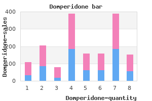
Joel C. Rosenfeld MD, MEd, FACS
- Associate Clinical Professor of Surgery, University of Pennsylvania School of
- Medicine, Philadelphia, Pennsylvania
- St. Luke? Hospital and Health Network,
- Bethlehem, Pennsylvania
Skin incisions Incisions must not cross flexor creases immediately medicine 6 year course buy domperidone 10mg online, as scar shortening will result in a flexion contracture of the joint medicine journal purchase domperidone 10mg visa. Tendon restore the strength of the repair is related principally to the variety of strands of core suture material that cross the injury medications memory loss best buy for domperidone, and to safe knot tying schedule 9 medications order discount domperidone. Rehabilitation the principal problems of tendon restore are re-rupture and tendon adhesion medicine that makes you poop order domperidone discount. Risk of these problems could be minimized by the adoption of an intensive supervised rehabilitation protocol that encourages early movement while protecting the restore with dorsal splintage that limits extension medicine 3604 order generic domperidone on-line. Nailbed Hyponychium of nail Phalanx (bone of fingertip) Dorsal view the brand new nail is formed). Obtain radiographs of the affected by way of the nail as a whitish, semicircular digit. The root of the nail Subungual haematoma: that is handled with is the deepest part, hid under the proxia trephine for symptomatic relief. The nailbed is sealed off underneath the affected digit with iodine or chlorhexifree fringe of the nail by the eponychium or dine. The release will current with a swollen, bruised fingertip of blood beneath stress is accompanied by that may reveal: relief of pain. Nail removing: this is carried out by pushing a small artery clip underneath the nail as far as fracture. A subungual haematoma has a normal harm to enable thorough lavage with sterile total appearance to the fingertip with dark saline. After vital nailbed laceration, which might lavage, pass a nice double-ended k-wire in an require surgical exploration, is commonly related anterograde direction by way of the distal with an avulsion of the nail root or a displaced Hand infections Avulsed nail root Subungual haematoma Avulsed nail 287 Nailbed damage 1 Nail removing and distally. Proximally, a suture is passed by way of the proximal nail fold, avoiding the germinal matrix, by way of the nail as a horizontal mattress, then again via the nail fold. Distally, a easy interrupted suture although the nail and underlying gentle tissue secures the nail plate in place. A piece of the aluminium foil packaging of a suture can be utilized in its place if the nail is misplaced or damaged. The old nail can be discarded after round four weeks as soon as the new nail has begun to grow. The cardinal features are a finger that is still flexed when the affected person makes an attempt to extend, but which then releases suddenly (like a trigger) and painfully with additional exertion or with passive manipulation of the finger. There is usually a palpable swelling of the flexor tendon on the stage of the A1 pulley, which could be felt shifting to and fro because the finger is moved passively. Patients should be referred non-urgently to the hand clinic, the place they might undergo an injection of steroid into the flexor sheath or be offered A1 pulley launch surgery. It might observe minor trauma, such as picking or biting at the nail or surrounding pores and skin, a minor penetrating wound, or waterlogging of the skin after immersion. Special care must be taken to keep away from inverting or everting the wound edges so as to prevent everlasting nail deformity. Nail plate alternative: the cleaned nail is then changed under the proximal nail fold A felon is an an infection of the fingertip pulp and as a dressing and spacer. It is held in place often follows a direct penetration similar to a with non-absorbable 5/0 sutures, proximally needlestick damage. An incision directly over the abscess with an eleven blade releases the abscess cavity. Fibrous septa inside the abscess cavity are broken down with a small artery clip to guarantee complete drainage. The finger is then dressed and the affected person is supplied with a short course of oral antibiotics. Incise the sheath, not only from the volar aspect of the sheath at each ends and send a sample of pus finger but in addition when the edges of the finger for microbiological investigations. Then insert a size 20 cannula into the proximal end of the are gently squeezed sheath and use it to irrigate by way of and three. Begin empirical intravenous Patients with suspected flexor sheath infec- antibiotics instantly after this lavage, rationtions must be referred urgently to Orthopaed- alizing them later according to the outcomes of ics or the local orthoplastic hand surgical procedure tradition and sensitivity analysis. This is a crucial prognosis as a outcome of flexor sheath infections usually require urgent surgical drainage, and uncared for infections are sophisticated by severe tendon adhesions and everlasting finger stiffness. The investigation and therapy of septic arthritis are described in Chapter 6 on page 108. Do not begin empirical antibiotics, as a bacteriology swab will be taken in theatre. Less extreme infections of small EmergencyDepartment interphalangeal joints may be managed nonmanagement operatively with elevation and systemic antiSeptic arthritis of the wrist or fingers presents biotics. Chondral harm is common and a with extreme ache at the joint, localized swelling, later joint fusion may be required. Spinal fractures are most likely to happen at zones of mechanical transition within the backbone; the thoracolumbar junction (T12�L2) is probably the most commonly fractured area, adopted by the craniocervical and cervicothoracic junctions. Pars interarticularis: these are the areas of the laminae that lie between the superior and inferior sides. Superior aspects: these bilateral projections lengthen superiorly from each pedicle and articulate with the inferior aspect of the vertebra above to type bilateral side joints. Inferior facets: these projections prolong inferiorly from the laminae and articulate with the superior facet of the vertebra below, on the aspect joint. Transverse processes: these lateral projections form the websites of attachment of muscular tissues and ligaments bilaterally. Anatomy Vertebrae Spinous processes: these project the vertebral column consists of 33 vertebrae: posteriorly within the midline and kind the 7 cervical, 12 thoracic, 5 lumbar, 5 sacral and bony prominences which are palpable four coccygeal. The parts of the vertebral arch comprise: Pedicles: these cylindrical projections join the body to the vertebral arch. Laminae: these sheets of bone are connected to the pedicles and coalesce at the base of the spinous course of to full the vertebral canal. The articulation between the skull (the occipital condyles) and the cervical spine is of the vertebral physique and disc. The axis has a peglike course of (referred the posterior column contains the poste to because the odontoid peg, or dens) that projects rior components. The odontoid peg Cervicalspine the primary two cervical vertebrae are signifi is held in place by two ligamentous constructions: cantly totally different in form from the remaining the alar ligaments, which emanate from the occipital condyles and insert into the tip of the five. General ideas Superior side C1 (articulates with the occipital condyles) 293 Odontoid peg Foramen transversarium C1 contains the cell bodies and synapses of the wire, while the white matter consists of the ascending and descending tracts that transmit sensory and motor data (the lengthy tracts). Knowledge of the anatomy of the long tracts helps in understanding and figuring out patterns of spinal wire injury. Sensory examination of both pain and proprioception is kind of specific to these two tracts: an abnormality in one or different sensation will find the crosssectional place of an incomplete spinal wire lesion, giving helpful prognostic info. This is as a outcome of gentle touch sensation ascends in both the ipsilateral posterior dorsal column and the contralateral anterior spinothalamic tract. Occipital Alar ligament condyle Transverse ligament C2 physique Posterior view with ligaments (vertebral arches removed) Anterior arch C1 Lateral mass C1 Odontoid Transverse peg ligament � the spinocerebellar tract transmits uncon Superior aspect Posterior arch Axial view scious proprioception that permits the coor dination of movement. The cell our bodies of the preganglionic nerves of the autonomic nervous system are situated within the subaxial cervical spine (C3�C7) consists of the spinal cord grey matter. This Spinalcord the spinal cord is made up of peripheral white leads to peripheral vasodilatation and hypoten matter and central grey matter. Biomechanicsofvertebral columninjury Vertebral column damage results from extreme pressure being utilized to the backbone. The magni tude, location and trajectory of this force can result in displacement with six degrees of freedom: 1. Movement on the stage of an unstable fracture can cause further neurological deficit, and so patients suspected of getting a spinal harm ought to be dealt with with the minimal amount of pressure and movement utilized to the spine. At the scene of damage, the affected person is immobilized with a spinal board, a rigid cervical collar, neck blocks and straps. Spinaltensionbands In flexion accidents, the posterior components act as a rigidity band. If they remain intact, the vertebral bodies will fail in compression (resulting in wedge and burst frac tures). Alternatively, the posterior components might fail in rigidity (distraction), by way of both the bone or the ligaments, resulting in a much more unstable fracture. Principles of management of major spinal trauma reflexes within the higher and lower limbs A compromised airway is protected with easy rectal examination, together with perianal and measures and using adjuncts (p. This includes cutting their clothes free whilst preserving warmth Breathing and dignity. Thoracic respiration actions could also be abol Logroll the patient to permit inspection of the ished or impaired after a cervical or thoracic again and to remove debris and clothing spine damage, and diaphragmatic respiratory could (Box14. Thoracic harm could affect the chest wall, lungs or heart and should Secondarysurvey Once immediately lifethreatening injuries have be assessed through the major survey. During this rigidity that occurs as the result of spinal twine part, a scientific examination of the neck and harm, and is characterized by low blood pres again is carried out. Neuro Positioning genic shock could be the solely indication of spinal the Emergency Department administration of twine damage in an unconscious affected person. Neuro childhood spinal accidents follows the same prin genic shock must be distinguished from ciples as for adults. A appropriately sized cervical spinal shock (a syndrome of twine dysfunction; collar, with a spinal board, sandbags and tape, is required. If a spinal injury or neuro logical deficit is recognized, an in depth peripheral Spinal cord damage with out radiological neurological examination is required. Neurological Neurological assessment within the acutely aware dysfunction can be delayed or progressive within the first few days. The cervical collar is taken off to allow examination of the neck and occiput, and the removing of clothes, particles and jewelry. One particular person controls the pinnacle, neck and airway, offering guide inline stabilization of the cervical spine and directing the manoeuvre. The fifth member of the group performs a whole examination, checking for: � signs of damage and abrasions � bruising � boggy swelling � palpable steps within the arrangement of the spinous processes, including uneven spacing between processes, or loss of longitudinal alignment in both the coronal or the sagittal aircraft � tenderness of the spinous processes assessed by a rectal examination. Examination the spine can only be cleared clinically in the following circumstances: � the patient should be fully alert and orientated. If radiographs are regular however the affected person continues to have moderate to extreme spine pain or tenderness, then the immobilization is left in place. An ambulant affected person who has not been immobilized is most conveniently examined sitting in a chair, with the examiner standing behind. Assess for: Clearing the backbone in the unconscious affected person Clearing the spine in unconscious sufferers stays controversial and local protocols must be consulted. Principles of administration of main spinal trauma 299 Radiologicalassessmentofthe injuredspine Cervicalspineseries Three radiographs are generally carried out: 1. Lateral view of the cervical backbone the entire cervical backbone from the occiput to T1 have to be shown. There are two corresponding measurements that point out the positioning of C1 on C2. The atlantodens interval anteriorly ought to be <3 mm in an grownup and <5 mm in a baby. Anterior soft tissue shadow: the prevertebral delicate tissues lie between the pharynx and trachea (which seem lucent) and the vertebral bodies. C5�C7: the delicate tissue must be no extra than one hundred pc of the width of the corresponding physique. Tip of spinous process line: Review the ideas of the processes for avulsion fractures. Note that the road passes through the C2 spinous course of, which is longer than the processes above and beneath. C3�C7 vertebrae the subaxial vertebrae should be assessed sequentially, tracing out the contour of each and checking the relationship of the joints above and below. C7/T1 junction the normal relationship of the cervical vertebrae ought to proceed into the thoracic spine, and this cervicothoracic relationship must be imaged in all suspected accidents. This is a skinny bone however the anterior arch can be seen clearly as a beanshaped, opaque ring mendacity anterior to the odontoid peg of C2. Increased soft tissue width is clear following cervical spine fracture with traumatic C5/6 spondylolisthesis, on this case a retrolisthesis, and formation of a retropharyngeal haematoma. This view permits evaluation of the odontoid peg and the relationship between the peg and the lateral lots of C1: � Trace the contour of the odontoid peg and search for lucent traces or tilting of the peg. B, the same radiograph with the side joints highlighted; note the common distribution and normal alignment. A, this lateral cervical spine radiograph is insufficient by itself, as it fails to show C7 and the cervicothoracic junction. Accurate identification of the spinal degree is possible by comparing the form and dimension of the spinous processes. Key points � Ten per cent of spinal fractures occur with a second spinal fracture at one other vertebral level. If one spinal fracture is recognized, careful consideration must be given to imaging the whole backbone. Principles of management of major spinal trauma Lateral thoracic/lumbar backbone 303 A B � Assess overall alignment by tracing the anterior and posterior margins of the verte brae and of the spinous processes. Compare the peak of the vertebral physique to that of the vertebrae above and beneath. Loss of peak of the posterior wall of the vertebral physique indicates the presence of a burst fracture. The alignment of the spinous processes must be assessed as for the cervical spine.
Diseases
- Cerebro facio articular syndrome
- Ti?che Jadassohn nevus
- Nicolaides Baraitser syndrome
- Anorexia nervosa
- X chromosome, trisomy Xpter Xq13
- Congenital facial diplegia
- Osteopathia striata cranial sclerosis
- Medrano Roldan syndrome
In the direct test symptoms torn rotator cuff cheap 10mg domperidone mastercard, the mutation causing the disease is the same as the one which alters the restriction website medications with dextromethorphan domperidone 10 mg without prescription. Its main limitation is that the disease-producing mutation(s) should be recognized if one is to test for them medications kidney disease purchase domperidone 10 mg on line. Carrier diagnosis in recessive illnesses Presymptomatic analysis for late-onset ailments Asymptomatic analysis for illnesses with reduced penetrance Prenatal prognosis Preimplantation testing Prenatal Genetic Diagnosis Prenatal prognosis is doubtless certainly one of the most common purposes of genetic analysis 4 medications walgreens cheap domperidone 10mg. Diagnosis of a genetic disease in a fetus might help in making an informed decision regarding being pregnant termination treatment renal cell carcinoma buy domperidone 10 mg online, and it typically aids mother and father in making ready emotionally and medically for the start of an affected baby medications xerostomia order 10 mg domperidone fast delivery. Fetal cells are present within the amniotic fluid and can be utilized to diagnose single-gene problems, chromosome abnormalities, and some biochemical disorders. The risk of fetal demise because of amniocentesis is estimated to be roughly 1/200. The villi are of fetal origin and thus provide a big sample of actively dividing fetal cells for prognosis. This method has the benefit of providing a analysis earlier within the pregnancy. Disadvantages are a better fetal mortality fee than with amniocentesis (about 1/100) and a small chance of diagnostic error due to placental mosaicism. The pedigree under reveals a family in which hemophilia A, an X-linked disorder, is segregating. A 22-year-old girl with Marfan syndrome, a dominant genetic disorder, is referred to a prenatal genetics clinic during her tenth week of pregnancy. A 66-year-old man (I-2) has just lately been identified with Huntington disease, a late-onset, autosomal dominant condition. Homozygosity for the normal allele (choice C) is inconsistent with the results proven on the gel. Choice E is wrong as a end result of Marfan is a dominant disease with no "service" status. Heteroplasmy (choice B) is related to mitochondrial pedigrees, and the phenylalanine hydroxylase gene is a nuclear one. In an X-linked sample, this may be characteristic of a female with two copies of the disease-producing allele and may be very rarely seen. Before her testing, he had a 50% chance of getting the disease-producing huntingtin allele. Fractures are described in terms of numerous characteristics: aetiology � the purpose for the fracture morphology � the fracture pattern severity � the diploma of comminution, or involvement of soppy tissue or a joint location � which part of the bone is affected displacement � how the bone fragments have moved in relation to one another. This radiograph demonstrates quite a few lytic lesions within the subtrochanteric regions of both femora. From Sproat C, Burke G, McGurk M: Essential these are the most common type of pathologi- Human Disease for Dentists (Churchill cal fracture, but are often considered sepa- Livingstone 2006) with permission. They most frequently happen as a outcome of osteoporosis, a progressive proximal humerus and backbone. Common sites of cause is vitamin D deficiency, typically ensuing osteoporotic fractures are the hip, wrist, from alcohol abuse or renal illness. The mechanism of injury is commonly a easy twist and fall, or a sporting accident, usually to the tibia, humerus or fingers. Spiral fractures could have butterfly fragments or comminution with increasing energy switch. They these fractures are attributable to a bending drive may result from explosive muscular contraction ensuing from a direct blow by a moving object. A torus (buckle) fracture happens when an applied force causes the compression facet of a bone to buckle. A greenstick fracture these fractures are brought on when bone fails in occurs if the strain side of the fracture also compression. These could be difficult damage to the knee), or at the humeral or femoral accidents to scale back. Physeal injuries warrant associated injury and problems, including particular consideration and are described on non-union. Neurovascular compromise is particularly essential and infrequently requires emergency treatment. The classification and administration of open and sophisticated fractures are described in Chapter three. Moreover, closed discount is commonly troublesome and the try can outcome in additional displacement. Open reduction and fracture stabilization are sometimes necessary, and may be required urgently in the presence of neurovascular compromise. Inversion injuries of the ankle generally end in sprains of the lateral collateral ligament complicated. The metaphysis consists of cancellous bone, has thin cortices and carries articular cartilage at the joint floor. It is tubular with thick cortices and is break up into thirds for descriptive purposes. Similarly, a reduction of a displaced fracture to a perfect place is termed an anatomical discount. A fracture (or an imperfect reduction) that is very practically anatomical is claimed to be minimally displaced. Fracture size: distraction/shortening shortening may be accompanied by kinking of angulation: varus/valgus in the coronal plane adjoining blood vessels, inflicting vascular comand flexion/extension within the sagittal plane promise. In the Emergency Department, these fractures require discount by manipulation or rotation: inside and external rotation translation: movement anteriorly, posteriorly, traction. A fracture with its apex pointing laterally is claimed to have lateral angulation or to be in varus. In the sagittal airplane, a fracture with its apex pointing posteriorly could accurately be stated to have posterior angulation, to be apex-posterior, or to lie in extension or recurvatum. The posterior side of the wrist is graphs of a protracted bone fracture should embody the termed dorsal, and the anterior aspect is joint above and the joint beneath. Translation occurs when the fracture surfaces have shifted sideways relative to each other. Rotation is often appreciated more fracture could also be stated to have lateral translation, simply on medical examination than on plain posterior translation, or each lateral and posteradiographs (or fluoroscopy). Many are eponymous and specific to a single harm sort, and the bone is first given a quantity: 1, humerus; in this e-book these are described within the sections 2, radius and ulna; 3, femur; four, tibia and related to that region. It offers a standard description of fracture kind is completely different for the metaphysis the principal patterns of diaphyseal and metaand the diaphysis. Orthopaedic terminology There are then three teams inside each Group 2 11 fracture kind: 1, 2 and 3, once more numbered in a hierarchy of increasing complexity. There are then three subgroups within each 1 A Simple 3 group: these are denoted by a decimal level and then three numbers, 1, 2 and 3. The kind and group codes are widespread to most fractures, are simple to decide to memory and are useful descriptors to use in routine scientific follow. The subgroups have poor inter- and intra-observer reliability and are principally tools for research. Spiral C Complex Bending Multifragmentary Within every fracture type, the teams point out growing complexity, with 1 indicating a spiral pattern, and a couple of and three rising comminution. Spiral Segmental Irregular this represents rising problem in reduction and fixation. Within sort B, these normally represent numerous sagittal and coronal fracture planes, though at both end of the tibia, the B varieties characterize differing degrees of impaction. Medicalanddrughistory A comprehensive enquiry should embody conditions that may alter any surgical plan, together with diabetes and anticoagulant medicine. Simple articular, simple metaphyseal Simple articular, advanced metaphyseal Complex articular, complex metaphyseal Socialhistory this should cover occupation, hand dominance, diploma of social independence, hobbies and sports activities, and, importantly, smoking and other medication. The administration of the unstable trauma patient is completely different and is described in Chapter 2. There Look Inspect the injured part before touching, wanting ought to be a history of a discrete occasion, and for: the exact description will often predict the type of damage current. For example, a blow Swelling: initially localized after harm but quickly become more diffuse. The location is important: search for seatbelt restraint patterns after a motorcar collision, or bruising within the sole of the foot indicating an necessary structural harm to the hind foot or midfoot. Abrasion: may be accompanied by ingrained thirteen dirt, which has to be eliminated to keep away from permanent tattoo marking. Deformity might embody a bony prominence or a defect within the contour of a bone, muscle or tendon. Gauge the extent of energetic motion (produced by the patient) and passive movement such as a knife or glass. You ought to assume (performed by the examiner) within the injured joint, that the wound has penetrated to bone until and above and below the level of damage. Frac- tion, a fractured bone, an avulsed or torn ture displacement in any aircraft might result tendon, or a neurological damage. Determine the presence and character of pulses distal to the harm and examine these to the uninjured A facet. Determine whether the motor and sensory supplies to the limb are intact and normal (Ch. Move Radiologicalassessment B Radiographs Good-quality radiographs are needed for enough evaluation. A, Bony tenderness arising from a fracture is painful on palpation of the bone from any path. Ultrasound that is commonly used to assess the integrity of soft tissues such because the Achilles and quadriceps tendons and the rotator cuff. In kids, uncertainties referring to the presence of physeal lines should normally be resolved by reference to an atlas somewhat than by taking extra radiographs. Traction movies are plain radiographs taken whilst traction is utilized distal to a fancy damage, typically permitting a better appreciation of the fracture configuration. Stress movies are additionally occasionally indicated to determine whether or not a joint is steady or unstable. Blood tests that is useful in the evaluation of suspected an infection or malignancy and infrequently required for the overall medical evaluation of the unwell patient or those about to endure surgical procedure. Principles of fracture management 15 Reduce the acceptability of a fracture place depends on the situation of the fracture, the degree of displacement present, and the practical calls for of the affected person. The acceptable limits of displacement are addressed in the region-specific chapters. Fracture reduction is achieved by both direct (open) or indirect (closed) means. Directreduction this implies surgical exposure, visualization of the fracture, and anatomical reduction of the key fragments. It is generally required for periarticular fractures where the congruity of the joint surface is essential. It is usually accompanied by compression and rigid fixation of the fragments, in the expectation of primary bone healing. Is the fracture place acceptable or does it Indirectreduction that is achieved closed (without exposing the fracture), and is achieved with traction and manipulation, which may be a brief lived intraoperative manoeuvre (such as when femoral nailing) or a definitive technique (such as when utilizing an exterior fixator). In periarticular fracture therapy, the applying of traction to a distal part of the limb leads to ligamentotaxis � the reduction of the joint by pressure on the encompassing ligaments. This is usually imperfect: for instance, on the wrist, the volar ligaments are shorter than the dorsal ligaments, and the fracture will usually be pulled into dorsal tilt. This may be resolved into the simple paradigm: Reduce�Hold�Move these factors are intimately related: the tactic employed to reduce a fracture will influence the choice of tips on how to maintain it (the fixation technique), and this in turn might be influenced by the importance of and problem anticipated in allowing the patient to move throughout subsequent rehabilitation. The former can typically be lowered closed, held satisfactorily in a forged for 4�6 weeks, and then rehabilitated relatively quickly and simply. Conversely, the pilon fracture can solely be decreased open, have to be held with absolute stability during therapeutic to ensure final alignment and joint congruence, and has to be rehabilitated slowly with extended restricted weight-bearing. Stability is clearly a spectrum, but fractures are often described in binary terms. They require assist only for the comfort of the patient and are normally best treated with removable splintage (orthoses), usually secured with Velcro. This permits the affected person to alter the gadget to enable for the development, and subsequent decision, of swelling. Move the timing of rehabilitation is a compromise between protecting the fracture discount through extended immobilization and avoidance of load-bearing, and avoiding joint stiffness, muscle wasting and impaired function by way of early motion. Around the canal is a collection of concentric lamellae or rings of bone matrix, embedded within which are the bone cells: osteocytes. Osteons are tightly organized and overlap, offering cortical bone with its high density. In the diaphysis of a protracted bone, a ring of cortical bone surrounds a medullary canal containing a central nutrient artery, bone marrow and cancellous bone. In the metaphysis, the cortex is skinny, and the architecture Medullary canal Unstablefractures Unstable fractures will displace with loading and require stabilization to prevent collapse. Fracture stabilization may be achieved nonoperatively with splintage or plaster, or surgically with using a selection of gadgets including plates, screws, nails and external fixators. The kind of stabilization offered will, in flip, affect the mechanical surroundings and the way the fracture heals. Covering the outer facet of the cortex is a layer of specialised connective tissue, the periosteum, which is extremely vascular and (unlike cortical bone) accommodates nociceptors. Both the periosteum and endosteum include progenitor cells that, after fracture, differentiate into chondroblasts and osteoblasts, which produce callus. Where strain of the tissue on the fracture web site is more than 10%, granulation tissue types; at between 2% and 10%, fibrous tissue forms; whereas, at under 2%, bone formation is feasible.
Many proteins undergo posttranslational modifications as they put together to assume their ultimate roles in the cell treatment gout discount domperidone 10mg. Important options of the genetic code embody: Each codon consists of three bases (triplet) treatment 4 stomach virus purchase domperidone now. Protein synthesis begins with methionine (Met) in eukaryotes medications on carry on luggage domperidone 10 mg on line, and formylmethionine (fmet) in prokaryotes medicine cabinet with lights buy cheap domperidone 10 mg line. For these amino acids having more than one codon 5 asa medications generic 10mg domperidone with mastercard, the first two bases within the codon are often the identical symptoms 39 weeks pregnant cheap domperidone generic. They also can cause modifications in enzyme activity, dietary requirements, antibiotic susceptibility, morphology, antigenicity, and lots of different properties of cells. A transition is a degree mutation that replaces a purine-pyrimidine base pair with a unique purine-pyrimidine base pair. A transversion is a degree mutation that replaces a purine-pyrimidine base pair with a pyrimidine-purine base pair. Crossover or recombination between homologous chromosomes is a traditional part of meiosis I that generates genetic variety in reproductive cells (egg and sperm), a largely helpful result. In a standard crossover occasion, the homologous maternal and paternal chromosomes change equivalent segments, and though the resultant chromosomes are mosaics of maternal and paternal alleles, no genetic data has been lost from either one. On rare events, a crossover may be unequal and one of the two homologs loses some of its genetic info. Cridu-chat (mental retardation, microcephaly, wide-set eyes, and a attribute kittenlike cry) results from a terminal deletion of the quick arm of chromosome 5. If a splice site is lost by way of mutation, spliceosomes could: Delete nucleotides from the adjoining exon. Mutations in splice websites have now been documented in many alternative illnesses, together with -thalassemia, Gaucher illness, and Tay-Sachs. A massive number of -globin mutations have been described, including gene deletions, mutations that sluggish the transcriptional process, and translational defects involving nonsense and frameshift mutations. A 9-month-old infant of Greek descent was delivered to the hospital by his parents because he became pale, listless, and frequently irritable. The attending doctor noted that the spleen was enlarged and that the infant was severely anemic. It is believed that, similar to sickle cell anemia and glucose-6-phosphate dehydrogenase deficiency, the abnormality of pink blood cells in -thalassemia might defend against malaria. Splenomegaly is due to the role of the spleen in clearing damaged purple cells from the bloodstream. The excessive exercise of the bone marrow produces bone deformities of the face and different areas. The most typical remedy is blood transfusions each 2�3 weeks, however iron overload is a serious consequence. Trinucleotide Repeat Expansion the mutant alleles in sure ailments, corresponding to Huntington disease, fragile X syndrome, and myotonic dystrophy, differ from their regular counterparts only within the number of tandem copies of a trinucleotide. In these diseases, the number of repeats typically increases with successive generations and correlates with increasing severity and lowering age of onset, a phenomenon referred to as anticipation. The normal protein accommodates five adjoining glutamine residues, whereas the proteins encoded by the disease-associated alleles have 30 or more adjoining glutamines. A major clinical manifestation of the trinucleotide repeat enlargement problems is neurodegeneration of particular neurons. The growth of the trinucleotide repeat in the mutant allele may be in a coding area or in an untranslated area of the gene. A summary of the important trinucleotide repeat expansion diseases is proven in Table I-4-2: Clinical Correlate Huntington disease, an autosomal dominant dysfunction, has a imply age-of-onset of 43�48 years. Symptoms appear progressively and worsen over a period of about 15 years till demise occurs. Mood disturbance, impaired reminiscence, and hyperreflexia are sometimes the first signs, followed by abnormal gait, chorea (loss of motor control), dystonia, dementia, and dysphagia. Cases of juvenile onset (<10 years old) are more severe and most regularly happen when the defective allele is inherited paternally. This bond will later provide energy to make a peptide bond linking the amino acid right into a protein. Because base pairing is involved, the orientation of this interplay might be complementary and antiparallel. Notice that the peptide bond forms between the carboxyl group of the amino acid (or growing peptide) in the P site and the amino group of the next amino acid within the A site. Formation of a Peptide Bond by a Ribosome During Translation Steps of Translation Translation happens in the cytoplasm of both prokaryotic (Pr) and eukaryotic (Eu) cells. In eukaryotes, translation fifty five Section I Molecular Biology and Biochemistry and transcription are fully separated in time and area with transcription in the nucleus and translation in the cytoplasm. The large subunit binds to the small subunit, forming the finished initiation complicated. There are 2 essential binding sites on the ribosome called the P website and the A web site. After formation of the first peptide bond, the P site is a binding site for the rising peptide chain. In the translocation step, the ribosome strikes precisely three nucleotides (one codon) alongside the message. Inhibitors of eukaryotic translation include cycloheximide and Diphtheria and Pseudomonas toxins. Chloramphenicol is a drug used to fight bacterial infections, including meningitis. If given to a newborn, nevertheless, this drug can trigger a probably lethal response. Generally, four levels of protein shape are distinguished: Primary-sequence of amino acids specified in the gene. Secondary-folding of the amino acid chain into an energetically stable structure. Some proteins, like collagen, contain neither but have their very own more attribute secondary buildings. Tertiary-positioning of the secondary buildings in relation to one another to generate higher-order three-dimensional shapes (the domains of the IgG molecule are examples). Tertiary structure additionally includes the shape of the protein as a whole (globular, fibrous). Tertiary constructions are stabilized by weak bonds (hydrogen, hydrophobic, ionic) and, in some proteins, sturdy, covalent disulfide bonds. Agents such as heat or urea disrupt tertiary construction to denature proteins, causing loss of function. Quaternary-in proteins similar to hemoglobin which have a number of sub- Blue lips, nail beds, and pores and skin (cyanosis) Death Low blood stress units, quaternary construction describes the interactions amongst subunits. There is a category of specialised proteins, chaperones, whose perform is to help on this process. Molecular chaperones operate in plenty of cell compartments, together with the endoplasmic reticulum, the place intensive protein synthesis occurs. Proteasomes and Ubiquitin Whenever protein synthesis happens in a cell, a number of copies of a specific protein could not fold correctly. These defective copies are covalently marked for destruction by the addition of a quantity of copies of ubiquitin. Degradation of Misfolded Proteins By Proteasomes Many proteins require indicators to ensure delivery to the suitable organelles. These proteins all require N-terminal hydrophobic signal sequences as part of their primary construction. N-glycosylation refers to the addition of sugar chains to the nitrogen of asparagine residues (N-linked). The N-linked sugar chain can further be modified upon entry in the Golgi (posttranslational modification). O-glycosylation refers to the addition of sugar chains to the hydroxyl group of both serine or threonine residues of the protein, and it occurs solely in the Golgi (posttranslational modification). Significantly, the structure and sequence of the oligosaccharide chains on proteins and lipids (glycolipids) are the idea of the A, B, O blood groups. Accumulation or ineffective focusing on of misfolded proteins Proteins synthesized in the endoplasmic reticulum must fold appropriately for transport to the Golgi and then to their final destinations. In sure genetic ailments, the mutation might cause all copies of the protein to fold incorrectly. This mutation causes the 1-antitrypsin protein to misfold and aggregate in the endoplastic reticulum, where it damages cells, eventually resulting in cirrhosis. Its perform is to shield cells by serving as an inhibitor of proteases launched throughout a traditional inflammatory response. Among the more than ninety allelic variants of the 1-antitrypsin gene, the Z and S variants are most frequently encountered with 1-antitrypsin deficiency. Most importantly, after they arrive in the Golgi apparatus, specific mannose residues positioned in their N-linked oligosaccharide chains are phosphorylated by N-acetylglucosamine-1 phosphotransferase, forming a critical mannose-6-phosphate in the oligosaccharide chain. This phosphorylation is the crucial event that removes them from the secretion pathway and directs them to lysosomes. Genetic defects affecting this phosphorylation produce I-cell illness by which lysosomal enzymes are released into the extracellular house, and inclusion our bodies accumulate in the cell, compromising its operate. Organelles whose main perform is to digest supplies that the cell has ingested by endocytosis. Contain a number of enzymes that, collectively, digest carbohydrates (glycosylases), lipids (lipases), and proteins (proteases). Especially prominent in cells such as neutrophils and macrophages, although they serve this essential function in virtually all cells. Major Symptoms of I-Cell Disease A youngster aged 5 months was referred to a specialist. Examination of the fibroblasts beneath the microscope revealed the presence of quite a few intracellular inclusions, which on electron microscopy were revealed to be giant lysosomes. Biochemical evaluation confirmed decreased levels of the lysosomal hydrolase �-glucuronidase throughout the fibroblasts, however elevated levels of this enzyme inside the culture medium. When a lysosomal enzyme is missing (for instance in a genetic illness corresponding to Tay-Sachs), the undigested substrate accumulates in the cell, usually resulting in serious penalties. It has a somewhat unique main construction in that a lot of its size is composed of a repeating tripeptide Gly-X-Y-Gly-X-Yetc. Three pro- chains assemble to type a triple helical structure (procollagen), which might now be transferred to the Golgi. The propeptides are cleaved from the ends of procollagen by proteases to type collagen molecules (also called tropocollagen). Like osteogenesis imperfecta, these syndromes are a result of locus heterogeneity by which defects in a quantity of different genes (loci) may end up in related signs. Characteristic options embody thin, translucent skin; arterial, intestinal, or uterine rupture; and simple bruising. A blood take a look at confirmed that the infant had low serum ceruloplasmin and solely 10% of normal serum copper levels. Common with Ehlers-Danlos illnesses, Menkes illness has a symptomology due, partially, to weak collagen. Consequently, an affected particular person could have extreme copper deficiency and all copper-requiring enzymes might be adversely affected. Lysyl oxidase requires copper and performs a direct function in collagen formation by catalyzing the cross-linking of collagen fibrils. A deficiency in the exercise of this enzyme and other copper-dependent enzymes can be immediately liable for the described symptoms on this toddler. For each mutation described within the questions below, choose probably the most carefully related sequence change within the options above. A nasopharyngeal swab obtained from a 4-month-old infant with rhinitis and paroxysmal coughing examined constructive upon culture for Bordetella pertussis. A 25-month-old Caucasian woman has coarse facial features and gingival hyperplasia and, at 2 months of age, began developing a number of, progressive signs of mental retardation, joint contractures, hepatomegaly, and cardiomegaly. Levels of lysosomal enzymes are elevated in her serum, and fibroblasts show phase-dense inclusions in the cytoplasm. Which of the next enzyme deficiencies is most according to these observations Golgi-associated phosphotransferase Lysosomal -1,4-glucosidase Endoplasmic reticulum�associated signal peptidase Cytoplasmic -1,4-phosphorylase Lysosomal hexosaminidase A 7. Parahemophilia is an autosomal recessive bleeding dysfunction characterised by a decreased plasma concentration of the Factor V blood coagulation protein. Deficiency arises from a 12 base-pair deletion within the Factor V gene that impairs the secretion of Factor V by hepatocytes and leads to an abnormal accumulation of immunoreactive Factor V antigen within the cytoplasm. Collagen, probably the most plentiful protein within the human body, is current in varying quantities in lots of tissues. If one wished to examine the collagen content of several tissues, one could measure their content of A. A 6-month-old toddler is seen within the emergency room with a fractured rib and subdural hematoma. Lysyl oxidase Prolyl hydroxylase -Glutamyl carboxylase Phosphotransferase in Golgi -1,4-glucosidase 10. Respiratory tract infections brought on by Pseudomonas aeruginosa are related to the secretion of exotoxin A by this organism. The nucleotide sequences of codons 506�511 in this region of the conventional and mutant alleles are in contrast below. Deletion of a phenylalanine residue causing a change within the C-terminal sequence D. Deletion of a leucine residue with no change in C-terminal sequence 70 Chapter four the Genetic Code, Mutations, and Translation 12. A 10-year-old boy with severe progressive skin ulceration, decreased resistance to infection, and impaired cognitive capacity has been diagnosed with a genetic deficiency of the enzyme prolidase. Frameshift mutation In-frame mutation Missense mutation Nonsense mutation Splice site mutation Answers 1.
Apply stirrup: the backslab is liable to alongside the shafts of the tibia and femur medications mexico domperidone 10mg on line, with further padding on the bony prominences of the ankle and fibular head treatment diabetes order domperidone 10mg, as well as the highest and bottom of the slab treatment yeast infection child order 10 mg domperidone with visa. This could be utilized as one giant medicine wheel wyoming buy domperidone 10 mg on line, U-shaped piece of plaster (twice the size of the backslab but solely four layers thick) medications list a-z order 10 mg domperidone free shipping, or as one stirrup to all sides symptoms bowel obstruction order generic domperidone on line. If there are any obvious weak points, further help can be provided by adding smaller pieces where essential. At the same time, verify that no areas of pores and skin are uncovered directly to plaster (which will otherwise trigger a thermal burn). Apply the bandage, being positive to cover the folded Full casts ends of the stockingette. Support the limb while the plaster cures to be sure that the ankle remains in impartial, and the knee at 30� of flexion. Perform safety checks: Check for complete seventy three padding between the casting material and the skin to avoid abrasions, especially behind the thigh and on the metatarsal heads. The most typical errors are to depart uncovered, tough plaster behind the thigh and to fail to support the metatarsal heads. Key level An essential element of tibial shaft fracture immobilization is upkeep of correct rotational alignment. As for backslab software, time is important, because the patient might be uncomfortable, the Reinforcementtechniques assistant(s) will gradually tire of holding the backslab might be tremendously strengthened by the limb, the discount might displace, and the additions that enhance its cross-sectional plaster will remedy inside a few minutes of wetting depth. Plaster roll method: Place the tail of the a clear understanding of the steps concerned in bandage towards the limb, with the bandage the process. Apply stockingette: A layer of stockingette is thickness, convey your hand underneath and spherical utilized to defend the pores and skin. Complete plaster: After every layer of nesses ought to be utilized by covering half bandage, easy down the floor to of the previous layer with each successive exclude trapped air and consolidate the turn. An further flip is required over the separate layers of bandage into one mass bony prominences at the wrist, and at each of plaster. Place a wanted over the shaft of the limb, and 4 small piece of wool padding across the or 5 to reinforce the wrist joint. Wet plaster: Hold the roll in a single hand and the tail in the different to stop it becoming misplaced in the mass of wet bandage. Remove the bandage and squeeze gently between your arms to take away extra water, keeping maintain of the tail. Roll the stockingette and wool back over the edge of the plaster before applying the last turn of the bandage at either finish in order that Full casts the rolled edge of the stockingette and wool present a neat and padded finish. Safety checks: Check that the edges of the seventy five Pressure utilized over a small area will lead to high strain under the plaster and skin compromise, so apply pressure with the heel of the hand and thenar eminence to create as broad an area as the dimensions of the bone will allow. Three points of pressure require three arms and an assistant is therefore important. Repeat radiographs: Obtain check radio- the flat of the hand to keep away from a pressure level. At the time of fracture, the periosteum at the the adequacy of three-point strain can be tension aspect of the fracture is torn, whereas the gauged from the ultimate radiographs by assessperiosteum at the compression side usually ing where the wool padding has been remains intact. Pressure is supplies, a solid can be immersed fully then applied on the dorsal facet of the wrist, in water. As the stress is utilized, the dorsal are therefore normally most well-liked within the fracture periosteum comes under rigidity, providing clinic setting. They generally mould much less well than plaster of Paris and the sides are more abrasive; application is due to this fact extra demanding. The wet casting material is usually extremely irritating to the pores and skin throughout software, and gloves must be worn routinely. If a collar-and-cuff has been supplied, this should be positioned to hold the hand and wrist above the level of the elbow; a dependent position should be averted. If a collar-and-cuff is used in these circumstances, the weight of the arm can Upperlimb cause discomfort. A broad arm sling helps Oedema and bruising are gravity-dependent the arm from elbow to wrist. Secure the loose apex of the triangle behind the point of the elbow with a safety pin. The collar-and-cuff this secures the hand able of elevation because it rests on the contralateral shoulder, and is used in hand injuries. Place a triangular bandage over the forearm and shoulder, with the apex towards the elbow and the hypotenuse hanging vertically. Place a triangular bandage beneath the forearm and shoulder, with the apex towards the elbow, and the hyponenuse hanging vertically. Tie string to the highest of the pillowcase after which attach it to a drip stand for support. This sling is extremely efficient however the patient may develop a sense of stiffness in the shoulder and require short periods of moveInpatients with injuries to the hand, wrist or ment outdoors the sling for comfort. The peak forearm can conveniently be treated with a of the drip stand can be modified to allow the Bradford elevation sling. Hold a pillowcase with its opening in the path of the leg at hip level will tend to result in the the ceiling. Place a triangular bandage over the forearm and shoulder, with the apex at the elbow and the hyponenuse hanging vertically. Where compromise is delicate, with modest swelling and a few reasonable discomfort, place the limb in high elevation and reassess after 15�20 minutes. The volar facet of the wrist and forearm, and the area over the anterior compartment of the leg are convenient. A completely into two sections with two longituplaster saw has an oscillating (not rotating) dinal noticed cuts) to release the limb completely. Do not slide the saw laterally in shallow cuts; the cutting motion must be up and down. Once the solid has been divided along its whole length, use plaster spreaders to separate the edges of the solid, and then cut all the padding and gentle dressings all the means down to skin along the entire size of the cast. Where there are surgical dressings or swabs that have hardened with blood clot, these also can trigger constriction and should be elevated carefully. Plastershears Plaster shears are inserted at one end of the cast between the casting materials and the wool, with the decrease handle placed parallel to the limb or maybe a little depressed. Orthoses with the decrease handle in order that the plaster fills the throat of the shears. Maintain a slight pushing force and perform the slicing motion with the higher handle, transferring it up and down like a beer pump. Use plaster spreaders to separate the sides of the solid, and then reduce all of the gentle dressings down to skin alongside the complete size of the cast. If the signs resolve rapidly, a crepe bandage is wrapped frivolously around the break up solid to prevent it from falling off, and to act as a splint. Possible diagnoses embody: compartment syndrome vessel entrapment arterial dissection. Assess looseness by trying to move the plaster proximally and distally, noting its tour in either path. Where the affected person has restricted motion, the cast ought to be trimmed or changed as acceptable. A recalcitrant complains of ache or pores and skin compromise, the ring can be coaxed off by compressing the forged should be eliminated or a window reduce in finger distally with twine and then easing the it to enable adequate examination. The aims of operative fracture management are to cut back and stabilize the fracture in order to restore the alignment of the limb, management pain, and permit early movement and rapid return of function. The advantages of accurate reduction and early rehabilitation should be weighed towards the inherent dangers of anaesthesia, surgical procedure and implantation of foreign materials. Successful fracture surgery demands a clear understanding of biomechanics and the impact of surgical intervention on the biology of fracture therapeutic. For periarticular injuries, traction is utilized via ligamentotaxis (tension on the periarticular ligaments and joint capsule), which permits some realignment of the joint. The degree of fracture stability achieved after inner fixation is described in phrases of two theoretical mechanical environments: absolute stability and relative stability. Absolute stability implies primary bone If a fracture needs to be decreased anatomically healing (without callus). An open reduction permits direct visible evaluation of the fracture, and the application of bone clamps that may make the reduction extra simple. An open reduc- Absolutestability tion is normally adopted by inner fixation with Absolute stability is achieved the place the fraccompression utilizing screws and plates, or a ture fragments are compressed and held rigidly tension band technique. It is commonly used folalignment, similar to within the therapy of most lowing the open anatomical discount and fixalong bone fractures. Compression is a achieved utilizing traction and closed manipula- key idea in attaining absolute stability and tion, and is assessed radiographically. However, if the opposite cortex is deficient, the moment arm will are inclined to trigger the assemble to collapse. External fixators are therefore normally inadequate for definitive stabilization of femoral or tibial fractures. The following are generally used: Screws Plates Fixed-angledevicesandlockedplates Tensionbandwire Externalfixators Intramedullarynails. An A-type transverse mid-shaft fracture of a protracted bone with interlocking interstices, which has been decreased anatomically, is intrinsically stable; when fixed with a well-fitting intramedullary nail, the bone and implant construct is highly stable. When the patient stands on this leg, a part of the physique weight is supported by the bone (through the fracture) and part through the nail. In contrast, a C-type comminuted fracture at the same stage has no intrinsic stability, and if the affected person have been allowed to take full weight immediately, the nail could be load-bearing. In between these extremes, a B-type fracture with a transverse space of contact will be load-sharing and stable after nailing, whereas a spiral or oblique B-type fracture could also be vulnerable to shortening initially. To shield the implant, full weight-bearing could additionally be delayed in kind B and C fractures till some callus has begun to form. The socket of the pinnacle has a selected form to accommodate a screwdriver, mostly a hexagon or a star. Note that, though the top of the head is flat, the under-surface is hemispherical. The separation of the threads is termed the pitch: this is the space advanced when the screw is turned as soon as. Cancellous screws could have thread alongside their whole size (fully threaded) or solely a bit at the tip (partially threaded). They might have a central cannulation that permits them to be placed over a information wire. A locking screw has threads in the head as properly as the shaft, allowing it to have interaction with specialised plates. Screws are generally referred to according to their outer diameter, the commonest sizes being three. In phrases of function, there are many makes use of to which a screw could additionally be put, regardless of its form. Blocking and locking screws are terms used close to intramedullary nailing. The drill size corresponds to the core diameter of the screw to be used (see Table 5. Place a faucet via the drill guide and use 4 it to minimize a threaded channel in the bone. This have to be launched during the together, the screw threads must grip one means of tapping by turning the tap antifragment solely, while the screw head pulls clockwise half a turn for every three clockdown on the opposite fragment. The most screws are now self-tapping and this diameter of the gliding gap is identical as step is often not necessary. Technical tip Inordinate quantities of time could be wasted in the course of the means of screw insertion when, in moving from one step to one other, an instrument fails to discover the right trajectory to move easily by way of the drill hole. Keep one instrument in the right trajectory always; thus when the drill is eliminated, maintain the drill information in place until the depth gauge is poised, able to be inserted. Then leave the depth gauge in place until the screw is mounted and again poised for insertion. Use the smaller-diameter drill bit (corre- sponding to the core diameter of the screw) to drill the far cortex. Use a countersink to cut a shallow groove in the cortex to accommodate the form of the screw head. A buttress plate is used to resist shear forces, usually on the Plates metaphyses, while a bridging plate is used to A plate is a device utilized to the outside of a span areas of comminution. The plate is first along side a lag screw, to management secured to one of the bone fragments; a central the shear or torsional forces between bone drill guide (with a green-coloured collar) is used fragments that might otherwise cause the lag to drill the pilot gap before a screw is inserted screw to fail. Attention is then turned to the opposite end of the plate and the opposite bone fragment. An offset drill guide (with a gold-coloured collar) is used to drill an eccentric drill hole (2). As the screw (termed a compression screw) is tightened, the top is pressured down the sloping contour of the hole and slides alongside the plate (3). The fragment of bone gripped by this screw is thereby pulled alongside the plate, compressing the fracture. Apply the plate as above, and as the compression screws are tightened, the far cortex is compressed first, and then the close to cortex, resulting in a good distribution of compressive drive throughout the fracture. This is must be positioned laterally or anteriorly quite prevented by using the plate benders to pre-bend than medially or posteriorly. Beware: if the plate is incorrectly placed on the outset to kind an obtuse angle (3), utilizing compression screws will solely lead to displacement (4). The Verbrugge clamp has two suggestions, certainly one of which is notched to grip a screw; the opposite is pointed and is placed inside a plate gap. The ingenious articulated compression system also requires a unicortical screw for buy, and may achieve substantial interfragmentary compression.
Cheap domperidone 10 mg overnight delivery. Zwangsstörung: Symptome Diagnose & Therapie | Die häufigsten Zwänge.