

Susanne Matthes-Martin, M.D.
- Associate Professor
- Head of Unit
- St. Anna Children? Hospital
- Stem Cell Transplant Unit
- Children?s Cancer Research Institute
- Vienna, Austria
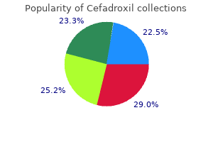
After this preliminary evaluation infection lung discount cefadroxil 250mg on line, checks should be undertaken to determine if the lady is ovulating and has patent fallopian tubes and if a semen sample of the male companion is normal antibiotics to treat diverticulitis cefadroxil 250 mg fast delivery. The bars symbolize possibilities calculated from information on 129 menstrual cycles in which sexual intercourse was recorded to have occurred on solely a single day in the course of the 6-day interval ending on the day of ovulation (day 0) antibiotics for uti and bv order 250 mg cefadroxil mastercard. The stable line shows daily probabilities primarily based on all 625 cycles prophylactic antibiotics for uti guidelines discount 250mg cefadroxil with mastercard, as estimated by the statistical mannequin treatment for recurrent uti in dogs cheap cefadroxil 250mg with mastercard. It is therefore thought-about optimum to carry out insemination or have sexual activity on the day earlier than ovulation antimicrobial resistance ppt order cefadroxil in india. Various vaginal lubricants and chemical compounds, as properly as saliva, used to enhance coital satisfaction could interfere with sperm transport. Some men experience midcycle impotence because of the pressure of performing intercourse on demand. Thus the phrases time and timing must be emphasized in the course of the preliminary counseling session. Couples also wants to be suggested to cease smoking cigarettes and consuming caffeinated drinks in excess. Cigarette smoking and caffeine consumption have been shown independently in several research to decrease the probabilities of conception. The common apply of vaginal douching additionally reduces the prospect of conception by roughly 30%. Preliminary information that the lady is ovulatory is supplied by a historical past of standard menstrual cycles. If the lady has common menstrual cycles, a serum progesterone level should be measured within the midluteal phase to provide indirect proof of ovulation in addition to regular luteal perform. Progesterone ranges of 10 ng/mL or larger are found throughout a minimal of 1 day of the luteal phase of normal ovulatory cycles during which conception occurred (Hull, 1982). Women with oligomenorrhea (menses at intervals of 35 days or longer) or amenorrhea who wish to conceive ought to be handled with agents that induce ovulation, no matter whether or not they have occasional ovulatory cycles. Therefore for these ladies, direct or indirect measurement of progesterone is pointless till after therapy is initiated. The endometrial biopsy is typically thought-about as a diagnostic methodology for the adequacy of ovulation and luteal operate. Abnormalities in the semen analysis (male factor) occurs in roughly 20% of couples with infertility as the sole issue and may be concerned in 30% to 40% of circumstances overall. The male companion must be suggested to abstain from ejaculation for 2 to 3 days earlier than assortment of the semen sample. It is best to collect the specimen in a clean (not necessarily sterile) wide-mouthed jar after masturbation. It is important that the complete specimen be collected, because the preliminary fraction incorporates the best density of sperm. Ideally, assortment ought to take place in the location the place the analysis might be carried out. The diploma of sperm motility must be decided as soon as possible after liquefaction, which usually occurs 15 to 20 minutes after ejaculation. Parameters used to evaluate the semen include quantity, viscosity, sperm density, sperm morphology, and sperm motility. The last parameter ought to be evaluated by means of share of total motile sperm in addition to quality of motility (rapidity of movement and amount of progressive motility). Sperm morphology is an extremely important parameter, which is correlated to fertilizing capability. Also, the semen profile displays sperm production that occurred three months earlier, which is essential to notice if there were sickness at that time. It is beyond our scope here to talk about absolutely the causes and diagnostic evaluation of semen abnormalities. When semen analyses have been performed on a bunch of men whose wives had conceived inside the past 4 months, approximately 75% had no less than one irregular attribute and 25% had two abnormalities. It is necessary to not miss a uncommon abnormality, corresponding to a testicular tumor; as nicely as it has been appreciated that male factor infertility is associated with different medical situations and subsequent problems (Eisenberg, 2015). Family historical past should be explored for genetically related sicknesses, start defects and, most importantly, the historical past of age of menopause in female members of the family. The bodily examination ought to give consideration to extremes of physique mass, pores and skin modifications, thyroid abnormalities, breast secretion, abnormal pain on stomach or pelvic examination, and evaluation of the vagina and cervix. In addition, if available, vaginal ultrasound performed at the similar time could also be extraordinarily Table 42. Increasingly it is suggested (although not mandatory) to display screen for genetic service status. Comprehensive screening for carrier standing together with Fragile X and different abnormalities corresponding to cystic fibrosis is easily carried out on the time of routine blood testing. Infectious disease screening (for chlamydia and gonorrhea) is carried out routinely in most practices at the time of the Pap smear. Levels are highest in young women and decrease with reproductive growing older; varied nomograms by age have been established (Seifer, 2011). In terms of ovarian reserve, greater levels (>2 ng/mL) counsel a bigger cohort of small available follicles and low levels (<0. Higher values, nonetheless, exhibit more variability in the early to midfollicular section. It is now established that use of oral contraceptive tablets decreases values by 15% to 20%. Use of Ultrasound in the Diagnostic Evaluation It is most typical to carry out a pelvic ultrasound analysis as a part of the investigation. By so doing, important pathology such as fibroids, endometriosis, and other pathology can be uncovered. Other Blood Testing Some specialists get hold of antibody titers for Chlamydia trachomatis, which if elevated might signify the potential of tubal illness. It has been suggested that if the immunoglobulin G (IgG) antibody titer is bigger than 1:32, 35% of sufferers have proof of tubal damage. Whether this sort of evaluation is routinely warranted as a focus for the infertility investigation continues to be debated. Provided that no other infertility elements are current, most anovulatory ladies (80%) conceive after induction of ovulation with therapeutic agents, and half the couples will conceive in the course of the first three ovulatory cycles (Gysler, 1982). As noted, most practices routinely display screen for chlamydia and gonorrhea during the preliminary examination. A water-soluble distinction medium permits higher visualization of the tubal mucosal folds and vaginal markings than an oil-based medium. It is important to be in a position to consider the appearance of the intratubal architecture to determine the extent of damage to the tube. The odds of being pregnant occurring after the procedure have been twofold higher when oil-soluble media had been used in contrast with water-soluble media. The procedure can even determine whether salpingitis isthmica nodosa is present within the interstitial portion of the oviduct. However, a diagnostic laparoscopy could also be thought of to detect the presence of peritubal adhesions. This could also be the outcome of uterine spasm (contractions) as a end result of the discomfort of the process, or due to true obstruction; the latter could also be only a light obstruction as a result of tubal particles. In common, the goal ought to be to have all tubal reconstruction carried out laparoscopically (discussed later). A suboptimal take a look at may be the results of approach, timing of the take a look at, and issues with cervical mucus or with sperm. In the past, this was an obligatory final step in the infertility investigation when all different check results were regular. Data have proven that in 20% to 40% of cases, some minor abnormalities 905 may be found. The probability that peritubal adhesions of enough severity to trigger infertility might be found on the time of laparoscopy is less than 5% in a lady with no history of salpingitis or symptoms of dysmenorrhea, a traditional bimanual pelvic examination, and normal antibody titers (if obtained) (Fatum, 2002). The analysis of luteal deficiency used to be made upon finding serum progesterone ranges persistently below 10 ng/mL 1 week before menses or discovering consistent evidence for a histologic delay (>3 days) within the pattern of the traditional secretory endometrium, indicating an insufficient impact of progesterone on the endometrium. B reveals injection of contrast by way of a 3-French catheter into the tube confirming profitable cannulation and a normal-appearing patent tube. There is at least a 10% disagreement of more than 2 days when the same observer dated the specimens on two separate events, and much more interobserver variability. As famous earlier, more modern studies have confirmed the lack of efficacy of the endometrial biopsy (Coutifaris, 2004). This essentially treats the luteal inadequacy, if it exists, preempting the need for invasive and imprecise endometrial biopsies. Substantial proof from animal research has indicated that antibodies could be induced in females from antigens obtained from organs within the male reproductive tract, and that these antibodies intrude with normal reproduction. Both spermagglutinating and sperm-immobilizing antibodies have been found in the serum of some infertile girls, but also within the serum of fertile management subjects. Agglutinating antibodies are discovered more frequently than immobilizing antibodies in most collection and, in some reviews, the incidence of sperm-agglutinating antibodies in infertile girls is just like that within the control group. Even with the finding of sperm agglutination or immobilization in interactions with serum (in vitro), it has not been demonstrated that an analogous diploma of sperm inactivation occurs within the decrease genital tract. One of the reasons for this discrepancy is that both serum assays measure mainly IgM and IgG antibodies, whereas the antibodies locally produced within the genital tract are primarily IgA. Thus some investigators have measured antisperm antibodies in cervical mucus and found a correlation between their presence and infertility. However, no information have shown that the discovering of antibodies towards sperm in the male or female partner is a reason for infertility. Autoimmunity to sperm in semen and serum has been found in some infertile males, significantly those that have had testicular infection, damage, or a surgical procedure corresponding to vasectomy reversal. Men with these antibodies have been treated with corticosteroid remedy and sperm-washing methods. Four prospective studies have reported the incidence of fertility occurring after a diagnostic infertility analysis was carried out in which the presence of antisperm antibodies was documented (Collins, 1993). These research were carried out in four completely different laboratories in three totally different international locations. All 4 research showed no correlation between the presence of antisperm antibodies in both member of the couple and the possibility of conception. Significance of Infectious Diseases in Subfertility Some researchers have instructed that asymptomatic, or occult, an infection of the higher female genital tract and male genital tract is a explanation for infertility. As early as 1973, it was suggested that infection with what was then called T-mycoplasma within the male might intrude with regular sperm operate, and an infection of the female reproductive tract could intervene with regular sperm transport. Two other microorganisms found in the feminine genital tract are Mycoplasma hominis and M. Although it has been reported that remedy of infertile couples with antibiotics, similar to tetracycline or doxycycline, that eradicate these organisms result in excessive being pregnant charges, managed research have reported no difference in being pregnant rates between couples treated with antibiotics and those not handled. Harrison and colleagues have studied 88 infertile couples with no demonstrable cause of infertility. One third have been treated with doxycycline, one third obtained placebo, and one third acquired no treatment. T-mycoplasma was isolated from approximately two thirds of the couples in every group and was eradicated only within the group treated with doxycycline. Nevertheless, conception charges had been related in each group (Harrison, 1975) as was additionally reported by Matthews and coworkers (Matthews, 1978) (Table 42. Other investigators have suggested that asymptomatic Chlamydia trachomatis infection may also cause infertility, but the dosage of doxycycline used in the randomized studies cited earlier would even have eradicated these organisms. In that the semen evaluation is subjective and variable, it has lengthy been advised that different more functional checks would enhance the analysis of the male partner (Oehninger, 2014). The zona-free hamster egg penetration take a look at initially described by Yanagimachi and associates was a take a look at developed to predict the fertilizing capacity of sperm and provides an additional, maybe more delicate, parameter for assessing sperm operate than routine semen evaluation. The sensitivity and specificity of the hamster egg penetration assay (sperm penetration assay) is taken into account to be too low to justify its routine use as part of the infertility investigation. Although these knowledge are older, this info from Hull nonetheless supplies one of the best out there comparisons. Among a group of infertile couples with unexplained infertility who were adopted for 2 years without remedy after the analysis was accomplished, it was found that the chances of becoming pregnant have been greater in girls younger than 35 years (75%) than in girls older than 35 (50%) (Hull, 1985). Rates for every age group are proven as strong squares, <25 years; blue triangles, 25 to 29 years; strong triangles, 30 to 34 years; blue squares, >35 years. In three of the 4 research of infertile couples who received no remedy talked about earlier, more than 50% of the couples who ultimately conceived did so within the first year after finishing the infertility evaluation. It has been instructed that certain couples have a better prognosis for spontaneous conception after the initial investigation, based on age and the time of making an attempt to conceive. This has been outlined as >30% inside a 12 months of the evaluation (Hunault, 2004), and it has been advised that in these couples, it is most likely not necessary to begin empiric therapy (discussed later). Generally, for couples presenting with the shortcoming to conceive, even if no factors have been discovered (unexplained infertility), in order to increase their probabilities of conception or to shorten the time interval till conception takes place, varied empiric remedies have been advocated. This typically increases the fecundity fee from a baseline of around 4% to 9% to 10% per cycle as might be mentioned later within the Unexplained Infertility section. Therapeutic brokers at present obtainable to induce ovulation are clomiphene citrate, letrozole, and urinary and recombinant gonadotropins. In addition, as mentioned in Chapter 39, if anovulation is attributable to hyperprolactinemia, dopamine agonists are an effective technique of inducing ovulation. As noted in Chapter 40, ovulation could also be induced by corticosteroid therapy in women with congenital adrenal hyperplasia. The former has a shorter half-life and is extra lively than the zu-clomiphene isomer, which has a for a lot longer half-life and is extra estrogen agonistic than antagonistic. The drug is usually given day by day for 5 days, beginning three to 5 days after the onset of spontaneous menses or withdrawal bleeding induced with a progestogen. Use of ovulation-inducing drugs alone has been shown to enhance the incidence of a number of gestations. Most start with an initial dosage of fifty mg/day for five days, starting on the fifth day of spontaneous or induced menses.

It is attributable to Gardnerella vagina/is antimicrobial insoles buy 250mg cefadroxil free shipping, a bacillus which usually grows when the vaginal flora shifts toward a extra acidic surroundings antibiotics pills buy cefadroxil american express. A watery antibacterial liquid soap purchase cefadroxil 250mg fast delivery, malodorous discharge without significant irritation is a standard symptom virus 1999 trailer purchase cefadroxil 250mg. Microscopically antibiotic misuse discount cefadroxil 250 mg without prescription, the bacteria overgrow and canopy the squamous cells antibiotics for acne cause weight gain order on line cefadroxil, producing so-called clue cells. Trichomoniasis, a sexually transmitted illness, is attributable to Trichomonas vagina/is, an oval protozoon with flagella. Microscopically, the organisms are recognized by their bluish-pink body, elongated nuclei, and flagella. Grossly, the virus causes a mucosal ulceration within a couple of days to 2 weeks following the publicity. These lesions are highly infectious till crusting, with last scarring occurring within 2 to three weeks of preliminary signs. Microscopically, the ulcerated lesions are characterized by epithelial necrosis with associated degenerated cells containing viral inclusions, finest recognized at the periphery of the ulcer. The cells with viral inclusions have attribute options including multinucleation and ground glass nuclei with a rim of chromatin condensation on the nuclear border surrounded by a cytoplasmic halo. Actinomyces-like organisms are most commonly seen in girls with noncopper intrauterine contraceptive gadgets. Atrophic vaginitis occurs mostly in postmenopausal ladies, but can also happen through the postpartum interval. Microscopically, the squamous cells present decreased glycogen as a result of lower estrogen ranges. Crohn illness can lead to rectovaginal fistula formation and is associated with fibrosis, continual irritation, and granulomas. The differential analysis contains vaginal fistulas of other etiologies including radiation remedy, perforated colonic diverticulum, or as a complication of hysterectomy. Stenosis, ulceration, and necrosis are well-described sequelae of radiation therapy. Stenosis can also follow severe bullous erythema multiforme (Stevens-Johnson syndrome). Epithelial inclusion cysts are lined by keratinizing squamous epithelium and full of white sebaceous and keratinous debris. Bartholin gland cysts are thought to develop from obstruction of the ducts of Bartholin glands, which usually open on to the vestibule. Adenosis normally includes the upper third of the vagina, but the middle third or lower third are affected in about 10% of cases. The diagnosis requires the presence of miillerian-type epithelium (most generally endometrioid) and endometrial-type stroma. Microscopically, squamous papillomas have a fibrovascular core and are lined by benign squamous epithelium. Fibroepithelial polyps mostly happen in grownup girls throughout their reproductive years. They happen in the decrease third of the vagina and grossly have a gentle and papillary floor. Leiomyoma is the most common benign mesenchymal tumor of the vagina in adults, with a imply age at presentation of forty years. Grossly, it consists of a well-circumscribed, firm mass with a white-tan cut floor. Microscopically, the tumor is composed of fascicles of spindle cells with elongated uniform nuclei, fine chromatin, easy nuclear membranes, and a average amount of eosinophilic cytoplasm. Genital rhabdomyoma is a rare tumor of the vagina that exhibits skeletal muscle differentiation. It affects middle-aged ladies, and patients usually current with vaginal bleeding or dyspareunia. Microscopically, rhabdomyoma is composed of loosely interweaving bundles of spindle cells with oval nuclei, abundant eosinophilic cytoplasm, and occasional crossstriations. Immunohistochemical stains for skeletal muscle markers corresponding to desmin, myogenin, and myo-D1 are optimistic. Rhabdomyoma could be distinguished from rhabdomyosarcoma on the premise of the absence of a dense layer of atypical neoplastic cells beneath the epithelium, cytologic atypia, and mitotic activity. Grossly, it has a well-circumscribed outline with a white-tan cut floor, and might range from zero. Architecturally, the cells kind alternating hyper- and hypocellular areas, with accentuation of the hypercellular areas around vessels. The absence of purple blood cell extravasation and stromal mucin distinguishes this entity from aggressive angiomyxoma. Deep 'aggressive" angiomyxoma predominantly impacts the sacroiliac delicate tissue and perineum of girls of their fifth decade. Postoperative spindle cell nodule is a pseudosarcomatous lesion that most commonly appears at the website of an excision, a couple of weeks to months after the surgical procedure. The differential diagnosis of postoperative spindle cell nodule includes vaginal leiomyosarcoma. It sometimes occurs in the upper vaginal wall of children with a imply age of 5 years. Microscopically, the squamous epithelium exhibits nuclear atypia (nuclear enlargement, hyperchromasia, and irregular nuclear membranes) with koilocytosis and an increased number of mitotic figures. Squamous cell carcinoma of the vagina accounts for 85% of vaginal carcinomas and occurs most commonly in women between the ages of 60 and 80 years. Squamous cell carcinoma typically metastasizes to the regional lymph nodes and has a predilection for distant metastasis to lung and bone. Microscopically, it demonstrates verruciform structure with minimal nuclear epithelial atypia, and a pushing somewhat than infiltrative margin. Patients sometimes current with bleeding or a grossly seen mass of the cervix or the vagma. Histologically, clear cell carcinoma is composed of cells with pleomorphic and hyperchromatic nuclei with plentiful clear cytoplasm; hobnailing is usually a outstanding characteristic. Primary adenocarcinoma of the vagina of nonclear-cell kind is rare; most non-dear-cell adenocarcinomas symbolize metastasis from the endocervix or endometrium, or different websites such as the ovary, colon, or breast. Sarcoma botryoides (a subtype of embryonal rhabdomyosarcoma) is the most typical malignant vaginal tumor in children, usually affecting girls <5 years. It is usually located submucosally and grossly appears as grapelike clusters of tumor that fill (and in some circumstances protrude from) the vagina. Microscopically, the tumor is composed of cells with elongated small nuclei and a reasonable quantity of bright eosinophilic cytoplasm. For prognosis, at least one microscopic field should present the malignant cells forming a condensed layer (a so-called cambium layer) beneath an intact epithelium (Pediatr Dev Pathol. The tumor cells are immunopositive for skeletal muscle markers such as actin, desmin, myo-D1, and myogenin. Surgical excision with radiation and chemotherapy is the treatment of choice, and the prognosis is usually wonderful. Current suggestions are that tumors >3 em in maximal dimension with an infiltrating margin, moderate to marked cytologic atypia, and;:::5 mitoses per 10 high-power fields be recognized as leiomyosarcoma (Obstet Gynecol. Microscopically, the cells have the same cytomorphology as the cells of cutaneous malignant melanoma. As famous above, metastasis often originates from malignancies of the cervix, endometrium, ovary, colon, and breast. The vulva or exterior female genital area encompasses the mons pubis, labia majora, labia minora, clitoris, and vestibule. The entire vulva apart from the vestibule is covered by keratinized, stratified squamous epithelium. The lateral aspects of the labia majora and the mons pubis include hair follicles. The clitoris is lined by keratinizing stratified squamous epithelium overlying paired corpora cavernosa that contain vascular spaces surrounded by nerves. Skene glands open on either facet of the urethral meatus and are composed of acini lined by mucus-secreting epithelium that open into ducts lined by transitional epithelium. Vulvar resections It is helpful to ask the surgeon to orient the specimen with a diagram or labeled sutures in order that orientation may be maintained throughout processing. The margins of resection should be inked and, relying on the location of the resection, the periurethral, vaginal, and perianal margins have to be famous. In instances with an apparent malignant neoplasm, one part per centimeter of tumor, together with the areas closest to the deep margin; lateral margin, and/or other margins are really helpful. In circumstances where no tumor is observed grossly, the whole specimen ought to be submitted. Because many gynecologic oncologists think about resection for squamous cancer in this space to be sufficient provided that tumor is >8 mm from the margin (Cancer. Many inflammatory and neoplastic conditions that affect the skin will also have an effect on the vulva. This section only covers these situations for which the vulva is a common website of disease. Bartholin abscess presents as a painful swelling in the space of the Bartholin gland. The etiology contains Neisseria gonorrhea, Staphylococcus, or other aerobic or anaerobic organisms. Hidradenitis suppurativa presents as painful subcutaneous nodules in areas containing apocrine glands, particularly the vulva and axilla. Initial adjustments *All e-figures are available on-line by way of the Solution Site Image Bank. Candida infection is commonly a persistent inflammatory condition of the vulva that might be related to diabetes. It typically presents as pruritis and clinically reveals areas of redness with thickened, edematous skin. Syphilis is a sexually transmitted disease attributable to the spirochete Treponema pallidum. The major lesion of syphilis, the chancre, develops in about half of the women within 3 weeks of infection and is characterized by one to sometimes a quantity of painless, dean-based ulcers. Secondary syphilis develops inside 6 weeks to 6 months and is characterised by the development of a rash on the palms, soles, and mucosal surfaces, as nicely as elevated plaques and papules (termed condyloma lata) on the vulva and mucosal surfaces. On microscopic sections, the chancre reveals epidermal ulceration, dermal acute and chronic irritation with quite a few plasma cells, and extreme arteritis. Condyloma lata are characterized by marked epidermal acanthosis and hyperkeratosis, dermal inflammation with numerous plasma cells, and arteritis. They current as asymptomatic, usually multiple or confluent, papillary or papular lesions, and may occur anyplace on the vulva or perianal region. Small condylomas could additionally be treated with topical agents whereas large ones are excised, or handled with laser ablation or cryotherapy. Molluscum contagiosum is a sexually transmitted illness in adults caused by an infection with the Molluscum contagiosum poxvirus. The lesions are small, 3 to 6 mm diameter papules with a characteristic central depression or umbilication, and are normally asymptomatic though perianal lesions could also be pruritic. Lichen sclerosus presents as symmetric plaque-like areas of white, thinned epithelium which might be superficially ulcerated. In advanced circumstances, there may be scarring of concerned areas and stenosis of the introitus. Lichen simplex chronicus (formerly "squamous cell hyperplasia") sometimes occurs in adults and presents as a localized area of pruritus (] Reprod Med. Clinically, the area is white or red, with accentuated skin markings and sometimes areas of excoriation. Treatment consists of limiting publicity to irritants, topical corticosteroids, and antipruritic brokers. Obstruction of the Bartholin duct results in the accumulation of secretions and the formation of a cystic dilatation of the duct. They are small, measuring only a few millimeters in maximal dimension, and are full of white tacky materials without hair. Mucus cysts happen within the vestibule and are lined by mucinous epithelium with or with out squamous metaplasia. They could also be hyperpigmented, hypopigmented, or flesh-colored, and usually occur on hair-bearing skin. They usually have a papillomatous or pedunculated progress pattern and a delicate minimize floor. Microscopically, the epithelium could additionally be thickened with hyperkeratosis, or could also be flattened. Papillary hidradenoma is a benign tumor that originates from apocrine sweat glands. It presents as a dome-shaped mass, usually <2 em in diameter, arising between the labium majus and labium minus. Microscopically, papillary hidradenoma types tubules and acini lined by a luminal layer of epithelial cells and an outer Clllptlr 315. Tumors may be exophytic, endophytic, or plaque-like and may be situated anyplace on the vulva. On microscopic examination, nests of invasive carcinoma will exhibit nuclear atypia, elevated mitotic activity, and will be associated with a reactive and desmoplastic stroma. In addition to the dimensions of the tumor, the thickness of invasive tumor (as measured from the top of the granular cell layer to the point of deepest invasion) in addition to the depth of invasion (as measured from the dermalepidermal junction at the tip of the closest regular dermal papilla to the point of deepest invasion) must be recorded. Lymphovascular area invasion is also an necessary prognostic feature and must be noted if discovered. The role of sentinel lymph node biopsy within the management of sufferers with vulvar squamous cell carcinoma continues to be evaluated (Curr Opin Obstet Gynecol. The intensity of the histopathologic analysis (including immunohistochemistry) determines the frequency at which metastases are identified, which is essential since even small metastases/isolated tumor cells are related to a small but increased risk for the presence of more extensive metastatic disease (Curr Opin Oncol.

The kind and variety of participants from whom biospecimens are collected depends upon the stated mission and out there resources of the biospecimen bank antimicrobial on air filters studies about order cefadroxil 250 mg with visa. In some cases bacteriophage order cefadroxil, small banks might acquire only defined specimens from participants enrolled in specific medical trials homeopathic antibiotics for sinus infection cheap cefadroxil online american express. In other circumstances antibiotic 93 3160 purchase cefadroxil on line, the bank rna y be disease-based and seek to generically acquire all obtainable tissue specimens for a given disease kind antibiotic eye drops for stye purchase cefadroxil 250mg otc. A corresponding informatics system is often required to observe and keep these information yeast infection 9dpo 250 mg cefadroxil free shipping. A biospecimen financial institution can also simply exist as a extra formal illustration of diagnostic paraffin block specimens already out there in the pathology division. Biospecimen banking is going to be a necessary ancillary activity to present tissue for emerging genomic, proteomic, and metabolomic testing methods. Several regulatory businesses have due to this fact produced tips for scientific biospecimen banks. Howevet; many elements of biospecimen banking are considered a research exercise and must conform to regulatory necessities that are completely different than for a medical laboratory. Policies have evolved considerably at the nationwide degree over the previous 5 years, and in addition range tremendously between states and establishments. In some circumstances, a Certificate of Confidentiality, a doc that asserts the best of the tissue financial institution director (or trustworthy broker) to protect the confidentiality of biospecimen knowledge even underneath court ordet; may be required (Genet Test. In most cases of prospective biospecimen collection, some type of informed participant consent is required. Explicit knowledgeable consent may be waived if it is inconceivable or impractical to acquire and the risk to the participant is minimal. Rarely, some institutions have dominated that generic language current in a hospital admissions doc or surgical consent form provides enough consent for biospecimen assortment, assuming that the specimens are distributed and utilized in a de-identified or nameless method. Generally, howevet; specific written consent for biospecimen banking should be obtained from the participant. Many generic templates for the language utilized in such a document can be found (] Clin Pathol. Since the principle danger to an individual participating in a biospecimen banking program is lack of confidentiality, measures used to defend such confidentiality and the dangers related to this loss are the principle dangers to convey to the participant. Howevet; when human biospecimens are getting used for genomic and disease-predisposition research, and when analysis results from biospecimen use generate patentable biomarker innovations, correctly documented extra particular informed participant consent may be important. Collected tissues may be snap-frozen or mounted in a variety of crosslinking or precipitating fixatives. Snap frozen tissue offers the very best high quality protein and nucleic acid derivatives, and is usually required for genomic or proteomic research. Howevet; the logistics of frozen tissue collection are tough and proper storage is comparatively costly. Fixation of tissue in precipitating fixatives such as ethanol, acetone, and others (Mod Pathol. They are the preferred biospecimen for proteomic analysis, but when correctly aliquoted from a number of sufferers from multiple time factors, can occupy a appreciable amount of storage space. Bone marrow may also be banked, particularly for analysis involving hematologic malignancies, other bone marrow dyscrasias, or occult tumor cell detection. However, large fluid quantity specimens (such as urine) may be difficult and costly to course of and retailer for lengthy durations of time, significantly for a future unknown use. Furthermore, evaluation of such biospecimens may require only a small amount of material per assay, which then necessitates that collections be excessively aliquoted to forestall multiple freeze/thaw cycles of the identical specimen. As an alternate, the cellular part of these fluids may be isolated by centrifugation and saved as cell pellets. Depending on the biospecimen kind and its supposed utility, there are a variety of necessary issues involved in collecting specimens for a biospecimen financial institution. For most general tissue financial institution actions, tissue specimens are collected in the center of routine remedy, corresponding to a surgical resection of a stable tumor, a therapeutic needle aspiration to take away joint or body cavity fluid, or a diagnostic bone marrow biopsy. In cases the place the diagnostic tissue specimen is limiting, properly supervised ex vivo sampling by core needle biopsy instrument or pores and skin punch may be used to acquire sufficient material for research without compromising the diagnostic specimen. Although few evidence-based guidelines exist, warm ischemia time must be limited and no less than documented in order to not introduce further preanalytical variability. Delays in blood processing can affect proteomic biomarker profiles (Expert Rev Proteomics. As with tissue, a quantity of business merchandise can be found to preserve blood cell elements at ambient temperature with out freezing, though these merchandise are comparatively costly and require specialised downstream isolation procedures (Cytometry A. For multi-institutional research protocols, it may be necessary to collect biospecimens from distant sites, typically from well being facilities or medical offices which have little expertise in dealing with intricate biospecimen necessities similar to snap-frozen tissue. Therefore, even restricted size biospecimens such as needle aspirates, tissue contact preps, and core biopsies are priceless and regularly amenable to molecular analyses. Obviously, nonetheless, such specimens should be judiciously distributed to maximize their analysis potential. For such circumstances, tissue specimens must be divided into samples no > 1 cm3 for fixation and/or freezing. Clinical and pathologic knowledge corresponding to the patient and specimen (for medical applications), and the participant and specimen (for research applications), are as necessary as the specimen itself. Without these related data, the utility of the specimens for clinical or correlative research is lost. The scope of the info which are collected and saved with the biospecimen will rely upon the mission of the biospecimen bank. It is often impractical to gather detailed medical info represented as written notes in a clinical chart or report; instead, it may be more efficient to rely on digital sources of data. Although information are often represented in textual content format, surgical pathology reports provide an correct and detailed supply of pathology info for tissue specimens that are collected and identified as a part of routine clinical care. In some centers, an digital medical record might provide entry to fundamental affected person demographics and scientific Chapter 62 � Biospecimen Banking I 9 21 diagnostic data. For most cancers patients and their specimens, the hospital tumor registry is often a fairly detailed and standardized supply for cancerrelated clinical and pathology knowledge. While smaller banks could depend on written log books and electronic spreadsheets to observe specimen information, these strategies become quickly constraining because the bank turns into bigger and extra diversified. Basic information sorts that might be required in a biospecimen financial institution data system embody the next: 1. For the lattet; the meant protocol should even be recorded, in addition to the consent underneath which the specimens had been collected, for the reason that consent probably regulates how the specimens could additionally be used. It is necessary to accurately keep the provision and storage location of each specimen in order that it might be rapidly retrieved on demand. Quality assurance measures corresponding to tissue histology evaluation, heat ischemia time, details of specimen processing, and nucleic acid high quality should be maintained for each specimen. As discussed above, a minimal set of clinical and pathology annotation knowledge should be related to every specimen. The efficacy of a biospecimen bank is judged nearly entirely upon its distribution of biospecimens for clinical testing or productive translational analysis. Therefore, detailed information regarding specimen distribution is essential in justifying the activity and operation of the bank. Several commercial software packages are available to assist in the administration of biospecimen banks. More frequently, individual centers develop their own information methods, often creating somewhat arbitrary and customized data schemes and knowledge definitions. While such applications may serve the quick wants of the financial institution, they present a significant obstacle to collaboration among different biospecimen useful resource centers. Among these instruments is the caTissue software utility, a freely available web-based software for managing biospecimen stock cabig. The identical principles of specimen processing used within the scientific laboratory apply to biospecimens banked for clinical and/or analysis purposes. Tissue may be frozen in liquid nitrogen, an isopentane cryobath (such as is available in most pathology frozen part rooms), or a make-shift dry ice ethanol bath. Any frozen storage system ought to be equipped with a temperature recording device to doc acceptable storage conditions and a remote alarm system that may contact the laboratory supervisor within the occasion of machine failure or energy loss. However, the standard of mounted tissue specimens may be improved using a number of quality control measures. By minimizing the time for which tissues are mounted, over-fixation (which results in excessive antigen crosslinking and molecular degradation) can be avoided. Documentation of processing times for every specimen can remove this preanalytical variable from tissue specimens collected over different time durations or from different places. Several commercially out there and "residence brew" precipitating fixatives protect tissue and tissue histology while minimizing the damaging effects to protein antigens and nucleic acids, brought on by cross-linking fixatives corresponding to formalin (Mod Pathol. The premise of this storage approach is that antigenicity and nucleic acids are shielded from additional degradation by both removal of oxygen and decreased temperature. For isolation and storage of serum and plasma, entire blood should be immediately spun and the suitable liquid element removed from cellular materials, aliquoted, and frozen. Processing must be carried out rapidly before cell lysis and protein degradation happens. Consortia of proteomics investigators have published suggestions on blood processing for proteomic research (Expert Rev Proteomics. For some types of genomic evaluation, it may be fascinating to have material derived from homogeneous cell populations. The ensuing recipient block can then be sectioned as a conventional tissue block, making it potential to carry out simultaneous immunohistochemistry or fluorescence in situ hybridization on every of the 300 cores on a single slide. Nucleic acid could be effectively derived from frozen and glued tissue using several standard protocols, although the quality from the later is usually inferim; as discussed above. Just as in any scientific laboratory, proper specimen labeling and identification are important, because a biospecimen bank could retailer biospecimens for extended intervals of time; maintenance of proper specimen identification is necessary. As biospecimen banks become topic to regulatory evaluation as with all clinical laboratory (as noted above), they might need to implement normal medical laboratory practices to forestall pattern mislabeling including use of preprinted barcode labels, double knowledge entry, and routine data auditing. They could also be collected at barely totally different times or areas, or might contain sampling bias. Such a evaluate of tissue or mobile samples might contain confirmation of a histopathologic diagnosis in addition to notation of basic mobile features corresponding to histologic preservation, tissue cellularity, and necrosis. As mentioned above, the quality of nucleic acid is directly related to the way during which the biospecimen was collected and preserved. Inherent necrosis and tissue cellularity in the specimen can even affect the standard of nucleic acids. Documentation of molecular specimen quality may be achieved by readily available (albeit often expensive) instrumentation designed for high quality evaluate of small amounts of nucleic acid, together with fiberoptic spectrophotometers and capillary microelectrophoresis methods. The success of any biospecimen financial institution is measured by the utilization of collected biospecimens. Distribution of biospecimens for patient care or funded analysis projects can additionally be an important income supply to support the larger biospecimen banking effort. Measures which may help promote the productive utilization of a biospecimen useful resource embody the next. Having a standardized and facilitated process for dealing with regulatory steps necessary for biospecimen banking and subsequent distribution will tremendously enhance utilization of a biospecimen useful resource. Clinical testing and translational research can only be accelerated when the biospecimens distributed are properly high quality managed and "assay ready. The strategy of case selection, specimen retrieval and processing, specimen annotation, and distribution can be timeconsuming and costly. Competing pursuits for limited biospecimens can also turn out to be problematic as financial institution utilization increases. Therefore, a biospecimen bank should set up a utilization evaluation committee so that the finite effort and physical resources of the financial institution could be fairly allotted to the scientific and analysis committees. These committees must be an unbiased illustration of Chapter 62 � Biospecimen Banking I 9 25 pathologists and scientists, with allocation decisions primarily based on medical necessity, scientific benefit of proposed tasks, investigator observe record with previous requests, funding standing, and probability that distributed biospecimens will meaningfully contribute to patient care or grant or scientific publication improvement. The prices may be borne by hospitals, since medical biobanking is part of the general hospital infrastructure required to support best affected person care. Clinical tissue banking may be compensated by way of establishment of particular billing codes that replicate the technical parts (specimen dealing with, high quality assurance, informatics, and storage) and skilled components (selection of appropriate tissue, microscopic review of banked specimens) of biospecimen banking. In return, the institution expects a return on its investment by method of the number of biospecimen-based projects, publications, and research grants that may be enabled by way of such a useful resource. Unlike traditional, hypothesis-based research grants, there are a quantity of extramural (noninstitutional) funding mechanisms to specifically support biospecimen banks. A viable specimen resource might want to develop investigator charges for the companies it supplies, including biospecimen collection, storage, and processing. These charges must be calculated realistically and bear in mind all operations and sources that are required from the initial collection of a biospecimen to its last distribution to an investigator. Such charges must be structured to additionally allow for financial institution development and additional development. Imaging Technologies in Surgical Pathology: Virtual Microscopy and Telepathology Jochen K. The future of surgical pathology will, nevertheless, include digital modes of image presentation to help diagnoses rendered by viewing the cells or tissue on a computer display screen rather than with an optical microscope, since digitized photographs of glass slides increase the clinically useful data that might be obtained from a pathologic specimen, and permit modes of research including digital consultations, morphometry, and slide storage that transcend the capabilities of present microscopy-based diagnostic pathology. The traits of the displayed picture, in terms of decision and vary of magnification, are primarily determined by the optical features of the scanning system and are total comparable with those of glass slide microscopy. However, in contrast to conventional gentle microscopy where the magnification, focus, and condenser setting can be adjusted at any time, for digital scanning, the "scanning depth" have to be determined previous to picture acquisition. The scanning depth consists of the area of curiosity, the scanning power, and the variety of horizontal ranges to be obtained in the plane of the tissue part (scanning energy contains the bodily magnification [e. Depth of focus is a requirement for interpretation of specimens that rely on a three-dimensional evaluation of mobile morphology, for instance, cytology specimens (Cancer Cytopathol. Unfortunately, no uniform (open-source) digital slide file format is presently obtainable, although interfaces are available that may convert picture information between the totally different digital slide formats. Scanning time, on the other hand, is still time-consuming, since scanning time is especially dependent on computational 926 Chapter 63 � Imaging Technologies in Surgical Pathology I 9 27 pace and optical physics, the latter of which is set by the magnification of the scanning goal. Some scanners shorten scanning time by using a meander rather than a linear scanning sample to purchase photographs, however each the meander and the linear methods have inherent physical limitations that turn out to be most apparent when acquiring multiple photographs inside each plane of section.
Agents used for bowel preparation might cause mucosal edema virus upper respiratory infection purchase cefadroxil 250 mg mastercard, hemorrhage infection years after root canal buy generic cefadroxil online, surface epithelial detachment antibiotic keflex buy generic cefadroxil pills, neutrophilic cryptitis antibiotic sinus infection order cheap cefadroxil on line, and increased apoptotic exercise antibiotics effective against strep throat buy 250 mg cefadroxil mastercard. So-called diaphragm disease within the small intestine is another instance of druginduced harm antibiotic resistant klebsiella uti purchase generic cefadroxil on-line. Histologic hallmarks in the intestinal tract embody a paucity of inflammatory cells in the lamina propria, apoptosis, and crypt dropout. Brown bowel syndrome, in contrast, is a singular situation believed to be attributable to vitamin E deficiency, which leads to mitochondrial dysfunction and lipofuscin accumulation in smooth muscle cells. Irritable bowel syndrome (ffiS) is a standard explanation for stomach ache and continual diarrhea. It is a medical prognosis, and colonic biopsies from these sufferers are completely normal on H&E stain. By definition, adenomas include at least low-grade dysplasia characterised by nuclear stratification; nuclear enlargement, elongation and hyperchromasia; and cytoplasmic mucin depletion. Paneth cells, neuroendocrine cells, and squamous cell clusters could happen in adenomas. Hamartomatous polyps can occur sporadically or as part of juvenile polyposis syndrome, Peutz-Jeghers syndrome, Cowden syndrome, and CronkhiteCanada syndrome. All these syndromes have an autosomal dominant inheritance sample, except for Cronkhite-Canada which is a nonhereditary dysfunction. Dilated crypts usually include neutrophils and/or mucin, hence the name "retention polyp. The polyps in sufferers with Cowden and Cronkhite-Canada syndromes carefully resemble juvenile polyps. It arises from the muscularis mucosae and presents as a well-demarcated nodular growth in the submucosa. Inflammatory polyp options inflamed lamina propria and damaged or distorted crypts, and granulation tissue could additionally be current. Inflammatory polyps may be microscopically indistinguishable from a juvenile polyp if cystic dilation of the crypts is outstanding; the suitable diagnosis depends on clinical information. Mucosal prolapse occurs most commonly within the rectum but could be seen anywhere in the colon (the alternative time period for this entity, solitary rectal ulcer, is a misnomer). These lesions are characterised by hyperplastic, tortuous crypts with plentiful irritation within the lamina propria. The distinction from well-differentiated adenocarcinoma lies within the lobular association of the displaced crypts, lack of dysplastic features, lack of desmoplasia, presence of surrounding lamina propria elements, and presence of hemorrhage or hemosiderin. Mucosal folds and prominent lymphoid follicles can resemble polyps endoscopically. Endometriosis, pseudolipomatosis, and xanthomas also can present as polypoid lesions. Acute appendicitis usually happens as the end result of luminal occlusion (such as by a fecalith, lymphoid hyperplasia, or Enterobius vermicularis), adopted by bacterial an infection. When inflammation extends into the mesoappendix and the serosa, periappendicitis should be diagnosed. Hyperplasia also can diffusely contain the appendiceal mucosa (mucosal hyperplasia). The lining epithelium is villous, serrated, and undulating and comprises a single layer of columnar or cuboidal cells with low-grade dysplasia. The associated mucin may be acellular, and lesions are principally associated with low-grade pseudomyxoma peritonei. Malignant glands invade the appendiceal wall and a desmoplastic response is current. Pseudomyxoma peritonei is primarily a medical diagnosis, and its prognosis is determined by the associated mucinous neoplasm. Although appendiceal lesions are answerable for the vast majority of the circumstances, mucinous tumors of other websites such because the ovary can not often be the trigger. Peritoneal mucinous carcinomatosis (high-grade pseudomyxoma peritonei features the presence of frankly malignant epithelium just like reasonably and poorly differentiated adenocarcinoma in a background of mucinous ascites. Neuroendocrine neoplasms are the most common neoplasms of the appendix and embrace a heterogeneous group of lesions with variable biologic conduct. Mesenteric fibromatosis is the commonest main tumor of the mesentery and most frequently happens in the mesentery of the small intestine. Histologically, spindle cells are dispersed in a densely collagenous stroma, and myxoid change of the stroma can be present. The borders of the lesion are infiltrative regardless of the well-circumscribed gross look. Patients with Gardner syndrome present comparable histologic findings, even though the myxoid stroma may be more prominent. Due to the infiltrative nature of the lesion, complete excision is tough, and recurrence is common. The scientific course is extra aggressive in Gardner syndrome patients; in reality, fibromatosis is the second most common cause of dying in this patient group. Sclerosing in mesenteritis is in the differential prognosis of mesenteric fibromatosis. The lesion is often solitary and most often arises from the mesentery of the small intestine, though in some instances diffuse involvement by multiple plenty may happen. Histologically, the lesion contains areas of fibrosis, irritation, and fat necrosis in various proportions. By immunohistochemistry, the tumor cells are smooth muscle actin-positive but nuclear beta-catenin-negative. A number of histologic patterns can be seen in several lesions or inside a single tumor. One frequent sample resembles nodular fasciitis with spindle to stellate formed cells embedded in a myxoid stroma. Other tumors show spindle cells organized in a storiform or a fascicular progress pattern. Prominent lymphoid aggregates, or lymphoplasmacytic infiltrates, are seen typically. Desmoplastic small spherical cell tumor is an aggressive malignancy mostly seen in younger boys and sufferers present with an abdominal or pelvic mass. The tumor consists of nests of small spherical blue cells embedded in a desmoplastic stroma separated by fibrous tissue. The tumor cells are vimentin and desmin optimistic; perinuclear keratin positivity can be present. The presenting symptoms are those of ischemia, thus the clinical differential analysis is usually quite broad. Idiopathic myointimal hyperplasia is a characteristic feature of the illness, consisting of a concentric proliferation of easy muscle cells throughout the small- to medium-sized veins of the affected mesenteric segment. The largest solid organ of the body, the mass of the adult liver is 1200 to 1600 g. Pressure equalization happens within the sinusoids, together with the nutrient and 02 gradient from portal tracts to terminal hepatic venules. The venous return is via the left, proper, and middle hepatic veins which be part of to form the inferior vena cava as it enters the guts at the proper atrium. Microscopically, the hepatic cords are lined by reticulin fibers and separated by sinusoids. The parenchyma is subdivided into acinar models of Rappaport which reflect an oxygen/nutrient gradient from most (zone 1) to least (zone 3), respectively; zone 2 is an ill-defined area in between. The lobule, usually used interchangeably with acinus, is a term based mostly on the idea of the hexagon by which hepatic cords radiate from the central vein toward the portal tracts. After measuring and description of the number of cores, liver biopsies are wrapped in lens paper and fixed overnight; use of sponge pads is strongly discouraged because of the artifacts created during sectioning. Protocol "special" stains and 6 extra unstained sections are really helpful for medical liver biopsies at preliminary preparation. Biopsies from immunocompromised patients (typically stable organ or bone marrow transplant patients) might or could not require "rush" processing; clear communication with the submitting clinicians is required in these cases and fixation, grossing, and processing are tailor-made to the clinical wants. Evaluation of intraoperatively encountered lesions is completed from recent tissue submitted on saline-moisturized gauze; cores are greatest sectioned with as little dealing with as potential, sectioned at ninety levels to the long axis. Frozen artifact creates areas that could be challenging to distinguish from fats; thus conservative estimates of the degree of steatosis are beneficial from frozen sections. If electron microscopic examination is anticipated for a potential metabolic illness, additional tissue should be fastened in 3% buffered glutaraldehyde. Iron and copper tissue quantitation can be performed in reference laboratories instantly from tissue in the paraffin block. The subcapsular regions are inclined to show parenchymal collapse; elastosis may occur in this location as properly as a outcome of continual ischemia. Wedge excisions for superficial, circumscribed lesions are managed similarly to resections, as described under. The surgical procedure might or might not follow anatomic boundaries, and thus before sectioning, it is essential to perceive the procedure that was carried out; evaluation of the imaging studies and stories is invaluable. After the type of surgical procedure, mass, and dimensions of the specimen are recorded, the resection margin is inked, the appearance of the capsule famous, and the specimen is sliced in the axial airplane at about zero. Gross examination of the lesion(s) and nonlesionalliver parenchyma ought to embrace color(s) (nutmeg; tan; bile-stained; hemorrhagic; yellow), viability (necrotic, nonnecrotic), texture (firm; exhausting; gentle; spongy), and presence of nodularity. Tumor sections (at least three) should reveal relationship of lesions to liver parenchyma, grossly visible vessels or ducts, margins (if close), and any variable areas within the tumor. Sections of nonneoplastic liver and inked resection margin(s) are submitted to consider underlying liver illness, vascular alterations, and margin status. One nontumor section of regular liver should be submitted and evaluated by routine special stains. Total hepatectomy (explant - carried out for end-stage persistent liver disease, fulminant hepatic failure, or metabolic disorders- is adopted by orthotopic liver transplantation. Radiology reports have to be consulted before processing the specimen to ensure that radiographically detected lesions are sampled. The hilum is completely removed, breadloafed, and submitted in toto proximally to distally with out dissection. The liver is then placed on the slicing board dealing with up, and sectioned axially cephalad-caudad in about zero. In their absence, two random sections from both the left and the proper lobes are submitted. In the strategy to liver biopsy, knowledge of the clinical info is essential. The adequacy of the biopsy must be assessed, which should be judged on the premise of the character of the question(s) being requested. Grading and staging schema had been initially developed for comparisons of treatment for autoimmune and "nonAnonB hepatitis" trials, however they shortly transitioned to apply to persistent hepatitis (Hepatology. Patterns of collagen deposition and architectural remodeling are frequently suggestive of the precedent damage: hepatitic, biliary, vascular, alcoholic, and so on. Viral hepatitis and alcoholic hepatitis differ as the previous is portal-based, and the latter is centered in zone three, is perisinusoidal initially, and leads to nodules the size of the acinus. The portal-portal fibrosis of the chronic biliary ailments often results in cirrhotic reworking with maintenance of the terminal hepatic venule in its central location; a "jig-saw" sample is subsequently suggestive of biliary disease. Acute viral hepatitis is commonly identified clinically by serologic and medical tests. Pathologists, subsequently, have comparatively little expertise with this type of liver disease. Histopathologically, acute viral hepatitis is characterised by simultaneous hepatocyte damage and regeneration, continual irritation, and no fibrosis. The changes embody swollen hepatocytes (hydropic degeneration), apoptotic (acidophil) our bodies, lobular spotty necrosis, lobular higher than portal irritation, sinusoidal cell reaction (Kupffer cell and endothelial cell hypertrophy), and bi- and multinucleated hepatocytes. The inflammatory infiltrates consist predominantly of mononuclear cells, with occasional plasma cells and eosinophils. Trichrome and reticulin stains may be complicated as the reticulin collapse may be in depth and the collapsed sinusoids might seemingly react with the stains for collagen. Fulminant hepatic failure or full decision might occur in any type of acute viral hepatitis. Plasma cells, macrophages, and eosinophils are also present, but in smaller numbe1. Some options are attribute of the particular forms of persistent viral hepatitis. Crystalline material is often current in substances that are abused by intravenous injection, and may be evident as polarizable materials inside portal macrophages. Adenovirus hepatitis, unusual in immunocompetent hosts and in adults, causes nonzonal foci of coagulative necrosis with a minimal inflammatory response. Bacterial infections contain the liver in numerous methods: space-occupying abscess, poisonous cholangitis. Sepsis may result in microabscesses, zone 3 canalicular cholestasis, and/or ductular cholestasis. Fungal infections are because of systemic infections corresponding to candidiasis, aspergillosis, and histoplasmosis. Parasitic infections, together with hydatid cyst, amebic abscess, and schistosomiasis, every with attribute histopathologic options, require a excessive index of medical and pathologic suspicion for prognosis. Steatosis, reversible with abstinence, is predominantly macrovesicular and initially involves zone 3. The prognosis is confirmed by attribute peripherallliC positivity, and serum electrophoresis phenotyping. Biliary atresia and Alagille syndromes are mimics, and it is essential to remember that globules may also occur in benign and malignant liver neoplasms. Excessive copper will not be visible by staining as it largely remains intracytosolic; diagnostic testing requires quantitation from the paraffin block (>250 1-Lg/g dry weight).
The shift toward an earlier diagnosis of breast cancer due to antibiotic bladder infection buy cefadroxil 250mg online improved imaging methods and screening applications highlights the need for biomarker discovery to quantify the residual danger of sufferers to point out the potential value of novel treatment strategies antimicrobial diet discount 250 mg cefadroxil amex. With the introduction of high-throughput technologies antibiotic resistance project buy discount cefadroxil 250mg, numerous multigene signatures have been identified that have the potential to outperform conventional markers (Endocr Relat Cancer antibiotics for dogs amoxicillin dosage purchase cefadroxil 250 mg without a prescription. Estrogen and progesterone receptors are parts of complex signaling pathways virus x aoba x trip purchase cefadroxil online from canada, which interact with a number of survival and proliferation pathways in the cell and play a critical role in the improvement and development of breast cancer antibiotic treatment for pneumonia purchase cefadroxil 250mg without a prescription. They have confirmed usefulness as prognostic factors, and extra importantly as predictive elements, in the medical administration of breast most cancers. There is growing evidence that sufferers with endocrine-responsive breast cancers benefit much less from adjuvant chemotherapy. Immunohistochemistry is the first-line attempt in evaluating hormone-receptor status, though it can be highly affected by a wide selection of preanalytic factors, together with time of tissue fixation and the antigen retrieval method(] Clin Oneal. In rare cases of solely PgR-positive tumors, the sufferers still profit from endocrine remedy. These quantitative assays have potential benefits in comparison with rnc strategies and may turn into the assays of selection in the future. Several studies have proven glorious concordance rate between analysis of biomarkers on needle core tissue and excisional specimen (Acta Oneal. In cases of unfavorable hormone receptors on core biopsy materials, reevaluation of the excisional specimen may be warranted to exclude a negative outcome due to tumor heterogeneity. To standardize immunohistochemical analysis of breast carcinoma, several scoring methods incorporating each intensity and proportion Chapter 18 � Breast Pathology I 321 of staining have been established. As a predictive marker, it has been related to responsiveness to anthracycline-based remedy and trastuzumab. As with hormone receptors, tissue dealing with, fixation, and processing can tremendously have an result on immunoreactivity of tissue samples. However, well-calibrated immunohistochemistry can determine nearly all of circumstances as optimistic or adverse. Recent expression profiling research of breast cancer have indicated the existence of no much less than 4 molecularly distinct types of breast cancer, which may originate from totally different cell types (Nature. Both luminal A and B cancers are inclined to reply to hormonal therapy, however luminal B cancers show a better response to chemotherapeutic brokers than luminal A. These outcomes are vital in the sense that they present, although breast most cancers exhibits important heterogeneity, from a biologic viewpoint many tumors may be categorised into explicit groups on the idea of genetic similarities, with gene signatures that correlate with clinical outcome and response to chemotherapy. On the idea of genomic profiling information, a quantity of genomic exams have been developed with the intent of providing even stronger prognostic information. The above assays can solely be performed by specific firms which have patented the test, and quality assurance have to be maintained throughout the company. Nonetheless, in present apply, histologic typing of breast tumors nonetheless remains the gold normal for classification of breast carcinoma. Although fats necrosis may end up from trauma, most cases are idiopathic, or are secondary prior to surgical procedure or radiotherapy. Duct ectasia usually presents in middle-aged ladies as pain and nipple discharge, with or without an associated mass lesion. Fibroadenoma is a quite common benign lesion of the breast that presents as a mass or radiographic abnormality, more generally in younger women. Grossly, fibroadenomas are grayish-white, firm, well-circumscribed, lobulated lots. Some fibroadenomas, especially these arising in the second decade of life, can grow rapidly and appear fairly mobile (an look that overlaps with benign phyllodes tumor); such lesions have been termed cellular fibroadenomas. Myxoid fibroadenomas display prominent myxoid adjustments within the stroma and barely may be a component of Carney syndrome (primary adrenocortical hypercortisolism, pores and skin hyperpigmentation, and a variety of nonendocrine and endocrine tumors). Fibroadenomas with significant epithelial proliferation have been Chapter 18 � Breast Pathology I three 25 termed complex fibroadenomas. Phyllodes tumor is much much less frequent than fibroadenoma, accounting for <3% of the fibroepitheliallesions. As talked about above, the main function distinguishing phyllodes tumor from fibroadenoma is the presence of the attribute leaf-like architectural pattern produced by intensive branching of the epithelial element. Stromal hypercellularity is the rule, usually with accentuation close to the epithelial clefts. Malignant phyllodes tumor reveals a distinguished infiltrative border, unequivocal sarcomatous areas, and stromal overgrowth (areas of stroma devoid of epithelium). Heterologous sarcomatous elements may also be occasionally present in malignant tumors. Overall, about 20% of the phyllodes tumors recur (ranging from 17% for benign tumors to 27% in malignant tumors) and 10% metastasize (0%, 4%, and 22% of benign, borderline, and malignant tumors, respectively). Phyllodes tumors are extraordinarily uncommon and their comprehensive diagnostic features could be discovered in more specialised texts. Peripheral papillomas present as incidental findings or sometimes as a mammographic abnormality. In the absence of any secondary neoplastic proliferation, major excision is an adequate therapy for papillomas. This relatively frequent lesion is usually incidental and admixed with proliferative lesions. However, it could current mammographically with calcification and/or architectural distortion, or clinically as a mass termed "adenosis tumor" or "nodular sclerosing adenosis. Elongated and compressed (as opposed to angulated) tubules, lack of a mobile desmoplastic stroma, and a lobulocentric pattern of development are useful diagnostic features. If doubtful, immunohistochemical demonstration of myoepithelial cells can moreover exclude an invasive process. In addition to their microscopic pseudoinfiltrative nature, these lesions may be clinically, radiologically, or grossly confused with invasive carcinoma due to their fibrotic nature and their attribute stellate/spiculated look. The presence of metastatic tumor deposits in axillary lymph nodes as determined microscopically is a highly unfavorable prognostic characteristic, as are a excessive variety of involved nodes and (to a lesser degree) large size of the deposits. Nodal standing (N) is so highly effective prognostically that it performs a significant role in determining remedy. The pathology report ought to include the entire variety of examined lymph nodes, the variety of positive lymph nodes, measurement of metastasis, and the presence or absence of extranodal extension by tumor deposits (the presence of extranodal extension is an indicator of tumor recurrence and its presence might dictate further radiation therapy). In all of those conditions, the size of the biggest deposit ought to be measured and reported. There are a couple of pitfalls within the analysis of axillary lymph nodes for metastatic carcinoma. The heterotopic tissue is subject to all changes that can happen in breast tissue in the mammary gland itself. The presence of myoepithelial cells and generally specialized stroma may be helpful diagnostic options. Ductal lavage has just lately been employed as a screening technique in women with a private historical past of breast cancer, however the sensitivity and accuracy of the strategy for detecting premalignant lesions of the breast ductal epithelium is still beneath investigation (Clin Lab Med. This prognosis is mostly rendered for benign breast lesions, together with inflammatory or infectious lesions, that are with out clinical or mammographic findings suspicious for malignancy. This analysis is rendered when the aspirate shows cells with worrisome cytologic options that fall in need of these required for a diagnosis of malignancy. This diagnosis is rendered when both the standard of the cytologic adjustments and the quantity of the malignant cells are adequate for an unequivocal analysis of malignancy. Fibroadenoma is a clinically well-circumscribed nodule with a homogeneous mammographic appearance. Gynecomastia is clinically a well-circumscribed and often painful subareolar lesion in a man. Fibrocystic changes usually current as a palpable, ill-defined lesion, which may have mammographically detectable microcalcifications. Subareolar abscess is a clinically painful, palpable subareolar mass typically associated with lactation. Ductal adenocarcinoma normally presents as a clinically palpable, mammographically suspicious mass, which cytologically exhibits a mobile smear containing large ductal cells, which maybe poorly cohesive, without a myoepithelial component. Lobular adenocarcinoma is probably not clinically palpable, however presents mammographically as an ill-defined mass lesion. Cytologic preparations show singly dispersed small plasmacytoid cells with vacuolated cytoplasm typically containing inspissated material. Immunocytologic analysis of estrogen and progesterone receptor studies can be carried out on cytospin slide preparations. World Health Organization Classification of Tumors: Tumors of Breast and Female Genital Organs. This chapter covers medical renal biopsies; biopsy for analysis of renal lots is covered in Chapter 20. In the basement membrane, electron-dense deposits may be subepithelial, subendothelial, or intramembranous (surrounded by basement membrane). Glomerular illnesses could also be major or secondary (associated with systemic diseases). Patients may be grouped into people who current with nephrotic syndrome (>3 g urine protein/day), nephritic syndrome (proteinuria+ hematuria), or isolated hematuria. It is necessary for the pathologist to have access to patient�s medical and laboratory knowledge. Diagnosis may be missed because of sampling error, particularly with the smaller needles presently used. The corticomedullary glomeruli are the primary to be sclerosed; due to this fact, needle biopsies should decide to pattern this area. Pathogenesis includes podocyte cell cycle dysregulation resulting in proliferation. Mesangial hypercellularity is defined as greater than three mesangial cells per glomerular section; many glomerular illnesses could have elevated mesangial cells (Table 19. Mesangial hypercellularity indicates mesangial immune deposits and/or reactive proliferation. Glomerular sclerosis is the most effective independent predictor of antagonistic consequence and renal failure. Both may recur in transplant kidneys, however clinical signs are gentle regardless of IgA deposition. Rarely, diffuse lgM deposits are detected (which have raised appreciable debate whether they symbolize a separate entity named lgM nephropathy). Other glomerulopathies characterised by isolated C3 or IgG mesangial deposits in sufferers without lupus stigmata have been described recently and named accordingly. Recent research have clarified the entity known as C3 glomerulopathy, which may present with mesangial hypercellularity or minimal adjustments. Interestingly, the illness is related to dysregulation of the alternative complement pathway secondary to defects in complement issue H (Nat Rev Nephrol. Most cases are idiopathic, but ""10% are associated with identifiable causes such as malignancy, autoimmune ailments. Glomerular lesions resemble those seen in Heymann nephritis, an animal mannequin by which antibodies react with the Heymann antigen, a fancy of megalin and Chapter 19 � Medical Diseases of the Kidney I 3 33 the receptor-associated protein. In Jones silver-stained sections, the basement membrane can show "spikes" projecting from the epithelial aspect of the basement membrane. As the illness progresses, deposition of basement membrane-like materials on the sides of the electron- dense deposits happens in order that with time, the electron-dense deposits are surrounded by basement membrane and thus turns into intramembranous. Occasionally, deposits are positioned predominantly in the mesangium (instead of in the loops) and are C3 or lgA/IgM instead of IgG (Semin Diagn Pathol. Additional morphologic findings are dependent on the sort of renal restricted or systemic disease. The medical prognosis of lupus is predicated on scientific and laboratory criteria established by the American Rheumatism Association. Renal involvement by the disease is comparatively frequent; approximately half of lupus patients develop lupus nephritis during the first year of the disease. The glomerular persistent lesions are segmental or global glomerulosclerosis, fibrous adhesions, and fibrous crescents. In the kidney, it will increase the propensity to pyelonephritis, papillary necrosis, arteriosclerosis, and glomerular disease. Classic X-linked (X-L) Alport syndrome presents with microscopic or gross hematuria and deafness. Renal biopsy is performed for prognosis as properly as evaluation of disease progression. Infectious brokers may end up in tubulointerstitial nephritis by colonizing the renal parenchyma, or by triggering a systemic immunologic response that can goal the renal tubules and interstitium. Most instances of acute pyelonephritis are ascending infections brought on by gram-negative bacteria, particularly Escherichia coli. When bacteria attain the kidney utilizing a hematogenous route, Staphylococcus aureus is normally the accountable agent. Acute pyelonephritis is characterised by an abundance of neutrophils in the lumen of tubules and the interstitium. Respiratory infection follows hematogenous spread, reaching organs just like the kidney, where it stays dormant; when the affected person later turns into immunosuppressed, the virus reactivates. It is useful to confirm the morphologic findings of viral an infection utilizing antibodies against the specific microorganism. A granulomatous inflammatory response is often seen with mycobacterial or some fungal infections. The histologic modifications are mainly confined to the tubules with necrosis of the lining epithelium. Epithelial cells can also exhibit regenerative modifications with nuclear enlargement, outstanding nucleoli, and mitotic exercise. Casts are often positioned in the collecting ducts, but as a result of retrograde filling, casts can even be seen within the proximal tubules. Casts are composed of sunshine chains and Tamm-Horsfall protein, and tend to have a fractured look. Nonspecific modifications similar to tubular atrophy and interstitial fibrosis are generally present. Vasculitides are also classified by the underlying pathogenetic mechanism as both immune-complex mediated or pauci-immune (absence of immune deposits).
Order cefadroxil 250 mg amex. SmartMouth Activated Oral Rinse - Dr. Clary.